Automatic segmentation method of brain gray matter nuclei in magnetic resonance imaging

A magnetic resonance imaging and automatic segmentation technology, applied in image analysis, image enhancement, image data processing, etc., can solve problems such as accurate and effective segmentation, affect diagnosis and research results, and cannot effectively distinguish nuclei, and achieve segmentation results precise effect

Active Publication Date: 2017-06-27
EAST CHINA NORMAL UNIV
View PDF3 Cites 6 Cited by
  • Summary
  • Abstract
  • Description
  • Claims
  • Application Information

AI Technical Summary

Problems solved by technology

[0003] However, due to the changeable morphology of these tissues, the currently commonly used atlas-based segmentation methods cannot achieve accurate and effective segmentation, and the results are quite different from the results of the gold standard segmentation map.
The level set segmentation method is a classic and effective automatic segmentation method, which can cope with variable tissue forms. However, due to the close space between these small nuclei located in the deep brain area, the classic level set method cannot effectively distinguish such as Substantia Nigra and Red Nucleus
This affects the results of diagnosis and research

Method used


Image

  • Automatic segmentation method of brain gray matter nuclei in magnetic resonance imaging
  • Automatic segmentation method of brain gray matter nuclei in magnetic resonance imaging
  • Automatic segmentation method of brain gray matter nuclei in magnetic resonance imaging

Examples


Embodiment

[0034] Segmentation of the red nucleus and substantia nigra of the brain's deep nuclei

[0035] The collected magnetic resonance image data is acquired by a multi-echo gradient echo sequence, and the data comes from a 3T magnetic resonance imaging equipment system (Siemens MAGNETOM Trio a Tim 3T), and the number of echoes used is 8.

[0036] The complex data collected by the gradient echo sequence undergoes steps such as phase fitting, phase unwrapping, background field removal, and a morphology-based dipole inversion algorithm (Morphology Enabled Dipole Inversion, MEDI) to reconstruct the brain transection magnetization rate graph.

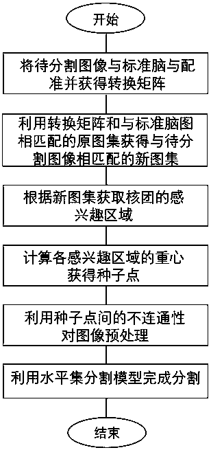
[0037] refer to figure 1 , is the flow chart of the present invention. After the quantitative magnetic susceptibility map is obtained by calculation, it is first necessary to register the calculated images of each layer with the standard template image. Using the same registration mode, the segmentation atlas corresponding to the standard templ...


PUM


Abstract

The present invention discloses an automatic segmentation method of brain gray matter nuclei in magnetic resonance imaging. The method comprises that by using standard reference images and a matched image set, seed spots located in each gray matter nucleus can be obtained through calculation; and a to-be-segmented image is preprocessed by using unconnectedness between the seed spots, fuzzy regions between adjacent nuclei are eliminated, and the segmentation contours of the target nuclei are automatically obtained by combining with the level set segmentation method. According to the method disclosed by the present invention, the variability of the morphology of the nuclei in different images can be more effectively coped with, and the segmentation results are more accurate; and by using the method disclosed by the present invention, specific nuclei such as the substantia nigra, the red nucleus and the like in the brain can be precisely segmented, diagnosis and pathological study on diseases such as Parkinson disease can be facilitated to a great extent.

Description

technical field [0001] The invention relates to the technical field of magnetic resonance imaging, in particular to a method for segmenting brain gray matter nuclei in magnetic resonance imaging. Background technique [0002] Magnetic resonance imaging technology has been widely used in the field of medical diagnosis. As an important means of diagnosis and treatment, its biggest feature is that it can provide rich image contrast information. With the continuous improvement of the spatial resolution of magnetic resonance imaging, magnetic resonance imaging technology can show the living fine gray matter tissue in the deep part of the human brain that cannot be observed before, such as the red nucleus, substantia nigra, dentate nucleus and some other located in the human brain. Brain nuclei of the basal ganglia, etc. Many studies have shown that these nuclei are closely related to some neurodegenerative diseases (such as Parkinson's disease and Wilson's disease), especially t...

Claims


Application Information

Patent Timeline
Patent Type & Authority Applications(China)
IPC IPC(8): G06T7/10G06T7/38G06T7/136
CPCG06T2207/10088G06T2207/30016
Inventor 郭天杨光李建奇薄斌仕
Owner EAST CHINA NORMAL UNIV
Why Eureka
Try Eureka