
Three-dimensional head testing method based on single camera module for radiotherapy

A radiotherapy, head three-dimensional technology, applied in the intersection of information science and radiation physics, the field of optics, can solve the problems of low speed, contradiction between precision and speed, poor precision and so on

Inactive Publication Date: 2017-07-18
SUZHOU WANGHAO INFORMATION TECH CO LTD
View PDF7 Cites 9 Cited by
  • Summary
  • Abstract
  • Description
  • Claims
  • Application Information

AI Technical Summary

Problems solved by technology

However, the current 3D inspection system has a contradiction between accuracy and speed
High-precision systems are slower; faster systems are less accurate

Method used


Image

  • Three-dimensional head testing method based on single camera module for radiotherapy
  • Three-dimensional head testing method based on single camera module for radiotherapy
  • Three-dimensional head testing method based on single camera module for radiotherapy

Examples


Embodiment 1

[0042] In the following, the invention will be described in a specific embodiment with reference to the accompanying drawings.

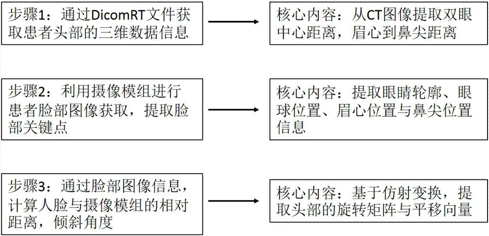
[0043] Step 1: Obtain a 3D CT scan image of the head from the patient's CT scan results before radiation therapy

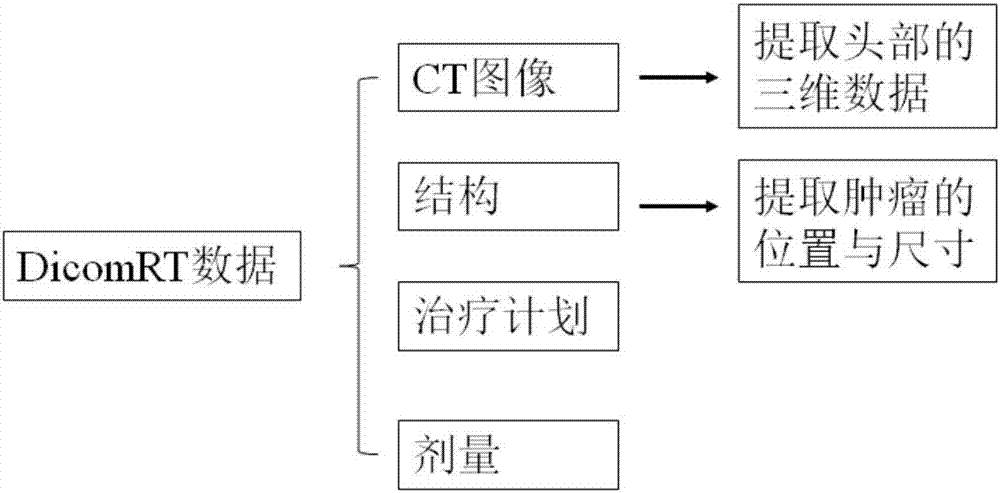
[0044] For cancer patients, careful diagnosis is required. Therefore, every cancer patient has corresponding 3D medical imaging data. At present, the DicomRT format is mainly used internationally to store cancer patient information. Its information structure is as figure 2 shown. The CT image is three-dimensional image data. The location and size information of the tumor is stored in the Structure data. Before radiotherapy, the DicomRT data will be obtained first, and the CT images and Structure data will be read from DicomRT according to its international standard coding rules. From the CT image, we can obtain the three-dimensional data information of the head, which includes the three-dimensional position information of each poi...


PUM


Abstract

The invention discloses a three-dimensional head testing method based on a single camera module for radiotherapy. The method comprises the following steps: step 1, acquiring a three-dimensional head image according to a CT scanning result of a patient before radiotherapy; step 2, acquiring a face image in real time during radiotherapy; step 3, calculating the current relative distance and the inclination angle between face and the camera module according tothe three-dimensional head image in the step 1 and the face image in the step 2; step 4, constructing three-dimensional coordinate points of a head in real time by taking the center of a camera sensor of the camera module as an original point. The face is shot by the single camera module, data of key points of the face is acquired, a rotation angle is obtained with an affine transformation algorithm, and a rotation matrix is obtained; a translation vector of the head can be obtained through combination with the three-dimensional CT image in DicomRT; three-dimensional positional information, including three-dimensional positional information of internal tumor, of the head in real space can be obtained in real time through actions of the rotation matrix and the translation vector on the three-dimensional CT image of the head, so that a tumor area is irradiated precisely by radioactive rays.

Description

technical field [0001] The invention belongs to the cross technical field of optics, information science and radiation physics, and specifically relates to a method of using a single camera module to detect the position of the key points of the human face in radiotherapy, and combining the three-dimensional tomographic images of CT to achieve the monitoring head The three-dimensional position of the tumor in the tumor, thus assisting the method of radiation therapy. Background technique [0002] Cancer has always been a persistent disease that threatens human health, even life. Cancer treatment methods mainly include surgery, chemotherapy and radiation therapy. Radiation therapy is a treatment that uses radiation to irradiate the tumor area. Radiation therapy is used in the treatment of about 70% of cancer patients, and about 40% of cancers can be cured by radiation therapy. Therefore, radiotherapy has an extremely important application value in the treatment of tumors. ...

Claims


Application Information

Patent Timeline
IPC IPC(8): A61B6/12A61B6/03
CPCA61B6/032A61B6/12A61B6/501A61B6/5247
Inventor 蔡夫鸿何赛灵
Owner SUZHOU WANGHAO INFORMATION TECH CO LTD
Learn More