Medical imaging method and device

A medical imaging and medical image technology, applied in the medical field, can solve problems such as PET image artifacts, achieve the effects of solving artifacts, improving image accuracy, and avoiding fusion deviation

Active Publication Date: 2017-08-04
SHANGHAI UNITED IMAGING HEALTHCARE
View PDF3 Cites 7 Cited by
  • Summary
  • Abstract
  • Description
  • Claims
  • Application Information

AI Technical Summary

Problems solved by technology

[0009] In view of this, the embodiment of the present invention provides a medical imaging method and device, which can solve the problem of PET image artifacts

Method used


Image

  • Medical imaging method and device
  • Medical imaging method and device
  • Medical imaging method and device

Examples


Embodiment Construction

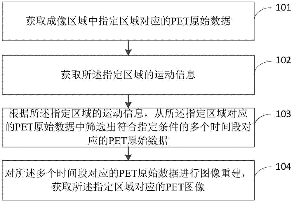
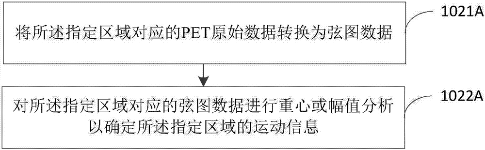
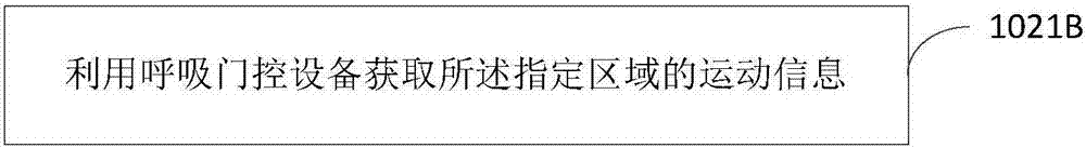
[0084] In order to better understand the technical solutions of the present invention, the embodiments of the present invention will be described in detail below in conjunction with the accompanying drawings.

[0085] It should be clear that the described embodiments are only some of the embodiments of the present invention, not all of them. Based on the embodiments of the present invention, all other embodiments obtained by persons of ordinary skill in the art without making creative efforts belong to the protection scope of the present invention.

[0086] Terms used in the embodiments of the present invention are only for the purpose of describing specific embodiments, and are not intended to limit the present invention. As used in the embodiments of the present invention and the appended claims, the singular forms "a", "said" and "the" are also intended to include the plural forms unless the context clearly indicates otherwise.

[0087] It should be understood that althoug...


PUM


Abstract

The embodiment of the invention provides a medical imaging method and a device, relates to the technical field of medicines and aims to solve the problem of PET image artifacts. The method comprises the following steps: firstly, acquiring a PET original data corresponding to an appointed area in an imaging area, and then acquiring motion information of the appointed area; screening the PET original data corresponding to multiple time periods that meet an appointed condition from the PET original data corresponding to the appointed area according to the motion information of the appointed area; and performing image reconstruction on the PET original data corresponding to multiple time periods, thereby acquiring a PET image corresponding to the appointed area. The technical scheme provided by the embodiment of the invention is suitable for medical imaging process.

Description

[0001] 【Technical field】 [0002] The invention relates to the field of medical technology, in particular to a medical imaging method and device. [0003] 【Background technique】 [0004] PET (positron emission tomography, positron emission tomography) is a display device that reflects the gene, metabolism and functional status of the disease. CT (computed tomography, computer X-ray tomography) uses X-rays to scan the human body. Due to the different density of tissues in various parts of the human body, the penetration of X-rays is affected, resulting in differences in the X-rays received by the scanning detector. , so that the lesion can be precisely located, and the fine structure changes of the lesion can be projected, but CT cannot reflect the physiological metabolism of the disease. [0005] PET-CT is an organic combination of PET and CT, using the same examination bed and sharing an image workstation, and combining PET images and CT images through image workstation proce...

Claims


Application Information

Patent Timeline
Patent Type & Authority Applications(China)
IPC IPC(8): G06T5/50G06T7/00
Inventor 顾笑悦孙友军刘伟平
Owner SHANGHAI UNITED IMAGING HEALTHCARE
Why Eureka
Try Eureka