Medicine carrying device for treating salpingostenochoria

A technology for eustachian tube stenosis, applied in the field of drug-loaded devices for the treatment of eustachian tube stenosis, which can solve problems such as bending and balloon dilation catheter breakage, and achieve the effects of reducing mucosal swelling, promoting regeneration, and smooth drainage

Active Publication Date: 2017-10-13
BIOVAS (WUHAN) MEDICAL TECH CO LTD
View PDF14 Cites 2 Cited by
  • Summary
  • Abstract
  • Description
  • Claims
  • Application Information

AI Technical Summary

Problems solved by technology

[0004] However, the end portion of the guide tube 3 of the above-mentioned special-purpose insertion instrument is a bent structure, while the existing balloon dilation catheter is arranged in a straight line. After the balloon dilation catheter is completely implanted into the guide tube 3, the balloon dilation catheter will Bending occurs due to the control of the lumen, and the control of the lumen can easily lead to the breakage of the balloon dilatation catheter

Method used


Image

  • Medicine carrying device for treating salpingostenochoria
  • Medicine carrying device for treating salpingostenochoria
  • Medicine carrying device for treating salpingostenochoria

Examples


Embodiment 1

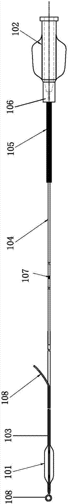
[0030] A drug-loaded device 100 for treating eustachian tube stenosis, comprising a first balloon body 101, a catheter body and a handle 102; the catheter body includes a distal tube body 103, a central hypotube 104, and a proximal tube body 105 ;

[0031] The first balloon body 101 is coated outside the end of the distal tube body 103; the left end of the central hypotube 104 is fixedly connected to the distal tube body 103, and the right end is fixedly connected to the proximal tube body 105; The proximal tube body 105 is connected to the handle 102; the connection between the handle 102 and the proximal tube body 105 is provided with a fixing part 106 matching a special insertion instrument.

[0032] As a preferred mode of this embodiment, the proximal tube 105 is a reinforced tube structure.

[0033] As a preferred manner of this embodiment, the central hypotube 104 is provided with an indicator mark 107 .

[0034] As a preferred mode of this embodiment, the distal tube ...

Embodiment 2

[0041] Repeat Example 1, the only difference is that the second balloon body 201 is only arranged on one side wall of the distal tube body, and after being filled with liquid filling medium, the distal tube body is bent to the other side Bending, matching with the bending section at the end of the guide tube of the special insertion device.

[0042] The working principle of the present invention is as follows:

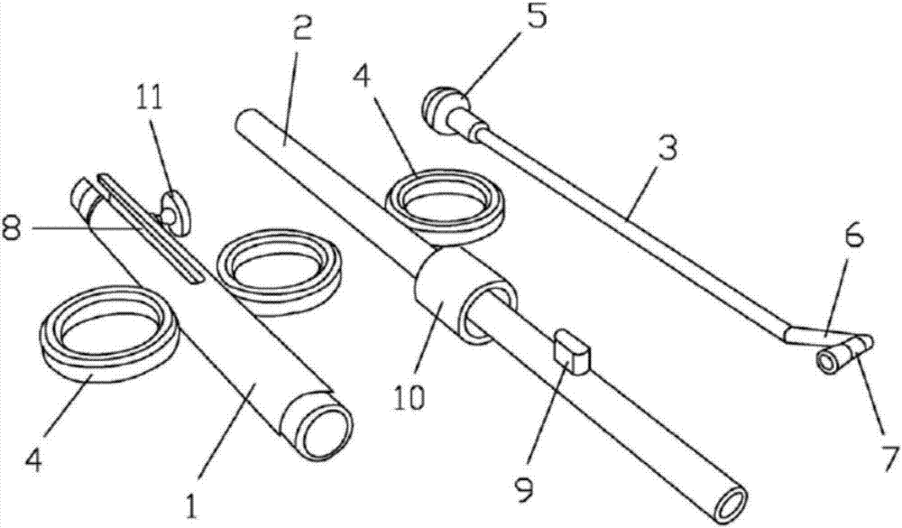
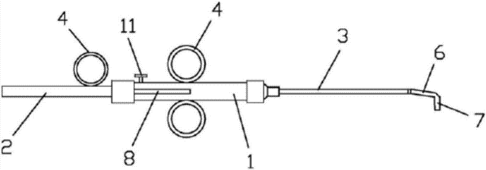
[0043] Main steps of the operation: Local anesthesia or general anesthesia can be selected. First, fix the knob 11, fix the push tube 2 at the distal position, and insert the drug-loaded device 100 through the push tube 2 into the guide tube 3. At this time, the balloon part It is completely hidden in the guide tube 3, and the drug-loaded device 100 is slowly pushed into the Eustachian tube along the guide tube 3. There should be no resistance during the push to avoid damage to the surface mucosa of the Eustachian tube. After entering the eustachian tube, connect the ...


PUM


Abstract

The invention discloses a medicine carrying device for treating salpingostenochoria. The medicine carrying device comprises a first balloon body, a catheter body and a handle. The catheter body comprises a far-end tube body, a middle wavy tube and a near-end tube body. The first balloon body wraps one end of the far-end tube body. The left end of the middle wavy tube is fixedly connected with the far-end tube body, and the right end of the middle wavy tube is fixedly connected with the near-end tube body. The near-end tube body is connected with the handle. A fixing part special for being matched with a built-in apparatus is arranged at the joint of the handle and the near-end tube body. A medicine coating is arranged on the surface of the first balloon body. The medicine carrying device can be used in cooperation with the medical special built-in apparatus, and operation is more standard; lumen is expanded after balloon dilatation, normal aeration of middle ears is restored, drainage is smooth, eustachian tube mucous membrane swelling is relieved, and meanwhile the regeneration and physiological function restoration of mucous membrane cilium are promoted; the problem that a balloon expanding catheter is prone to being broken in a catheter of a special built-in apparatus is solved.

Description

technical field [0001] The invention relates to the technical field of medical devices, in particular to a drug-loading device for treating eustachian tube stenosis. Background technique [0002] The Eustachian tube connects the middle ear cavity with the nasopharynx. Its tympanic orifice opens to the front wall of the tympanic cavity, and the pharyngeal orifice opens to the side wall of the nasopharynx. An important factor in sound transmission. For a type of middle ear disease caused by mechanical obstruction of the eustachian tube, such as adhesive otitis media and refractory secretory otitis media, etc., the conventional method is to perform puncture, incision or tube placement of the tympanic membrane. On the one hand, this method will damage the tympanic membrane, Tympanic adhesions do not improve significantly after extubation, or tympanic effusions are likely to occur repeatedly. On the other hand, there may be residual tympanic membrane perforation that requires se...

Claims


Application Information

Patent Timeline
Patent Type & Authority Applications(China)
IPC IPC(8): A61F11/00A61M31/00
CPCA61M31/00A61F11/00A61M2210/0662A61F11/202Y02A40/81
Inventor 陶忠许平董成旭王昊飞王海军
Owner BIOVAS (WUHAN) MEDICAL TECH CO LTD
Why Eureka
Try Eureka