Syringe needle with hemostatic function and preparation method thereof

The technology of a syringe and a needle tube is applied in the field of a syringe needle and its preparation, which can solve the problems of prolonged bleeding time and the like, and achieve the effects of good dispersion, uniform particle size and strong adhesion performance.

Inactive Publication Date: 2017-11-21
AFFILIATED HOSPITAL OF NANTONG UNIV
View PDF6 Cites 7 Cited by
  • Summary
  • Abstract
  • Description
  • Claims
  • Application Information

AI Technical Summary

Problems solved by technology

For patients with blood coagulation disorders, such as hemophilia, diabetes, or advanced cancer, the bleeding time after acupuncture is significantly prolonged, so c

Method used


Image

  • Syringe needle with hemostatic function and preparation method thereof
  • Syringe needle with hemostatic function and preparation method thereof
  • Syringe needle with hemostatic function and preparation method thereof

Examples


Embodiment 1

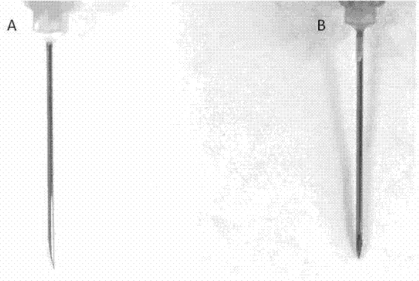
[0047] Preparation of syringe needles with polydopamine coating

[0048] First weigh 0.121g of tris(Tris, Mw =121.14) and add ddH20 to fully dissolve it, then adjust the pH value to about 8.3 with HCl, and finally set the volume to 100 mL to prepare a concentration of 0.01M Tris-HCl Weak alkaline buffer. Then 200 mg dopamine (dopamine, (4-(2-aminoethyl)benzene-1,2-diol), Mw=153) was weighed and added to 100 mL 0.01M Tris-HCl weak alkaline buffer to fully dissolve to obtain 2 mg / mL dopamine solution, then soak the needle tube of the syringe into the dopamine solution, stir at room temperature, make the dopamine slowly oxidize and self-polymerize, and form a layer of polydopamine coating on the surface of the needle tube in about 3 hours, such as figure 1 shown, figure 1 It is the syringe needle tube coated with polydopamine coating provided by Example 1 of the present invention.

Embodiment 2

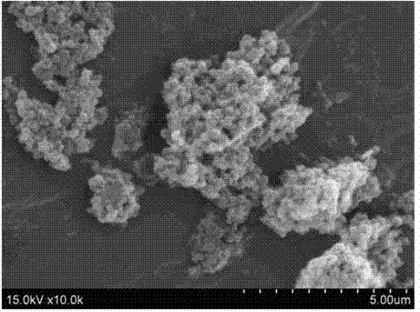
[0050] Preparation of chitosan nanospheres

[0051] Weigh 100 mg of chitosan and dissolve it in 50 mL of 2% (volume ratio) acetic acid solution, add 0.5 mL of nonionic surfactant Tween-80, stir on a magnetic stirrer at a speed of 500 r / s, and then While stirring, 10 mL of 1 mg / mL sodium tripolyphosphate solution was slowly added dropwise for crosslinking to obtain a chitosan nanosphere solution, which was centrifuged, and the precipitate was washed with water to obtain a chitosan nanosphere solution. like figure 2 , it can be observed that the chitosan nano-microspheres are spherical and uniform in size by scanning electron microscopy, and the particle size distribution of the above-mentioned chitosan nano-microspheres is measured by dynamic light scattering. figure 2 It can be seen that the particle size distribution of nano-microspheres is in the range of 110-320 nm.

Embodiment 3

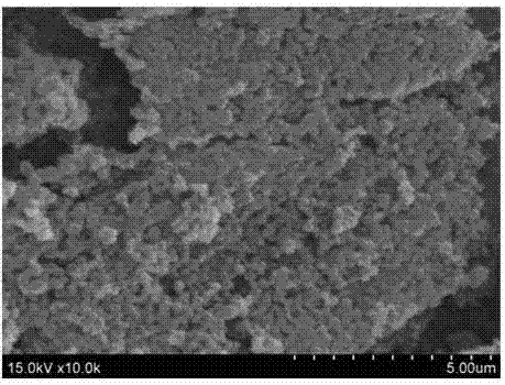
[0053] Preparation of Chitosan Nanospheres Loaded with Hydrophilic Drug Norepinephrine

[0054] Weigh 100 mg of chitosan and dissolve it in 50 mL of 2% (volume ratio) acetic acid solution, add 0.5 mL of nonionic surfactant Tween-80, then add 0.5 mL of 1 mg norepinephrine solution, and place on a magnetic stirrer Stir at a speed of 500 r / s, then slowly add 10 mL of 1 mg / mL sodium tripolyphosphate solution dropwise while stirring for cross-linking to obtain a chitosan nanosphere solution, centrifuge, and wash the precipitate with water. A chitosan nanosphere solution was prepared. like image 3 , it can be observed that the chitosan nano-microspheres are spherical and uniform in size by scanning electron microscopy, and the particle size distribution of the above-mentioned chitosan nano-microspheres is measured by dynamic light scattering. image 3 It can be seen that the particle size distribution of nano-microspheres is in the range of 140-330 nm.


PUM

PropertyMeasurementUnit
Particle sizeaaaaaaaaaa

Abstract

The invention discloses a syringe needle with a hemostatic function and a preparation method thereof. The syringe needle comprises a syringe needle main body, and chitosan nanospheres and/or drug-loading chitosan nanospheres for promoting a hemostatic function and coating the outer surface of the syringe needle main body. The polymer material such as polydopamine uniformly bonds the chitosan nanospheres to the surface of the syringe needle under action of a certain adhesion force. The syringe needle is used for in-situ solid-gel phase transfer after clinical tissue puncturing and needle withdrawal so that the perforation tissue is sealed and thus the purpose of rapid bleeding is realized.

Description

technical field [0001] The invention relates to a syringe needle tube with a hemostatic function and a preparation method thereof. Background technique [0002] Clinical tissue puncture with a syringe needle is often used to provide drugs and blood sampling for medical diagnosis, etc., and bleeding after needle removal is largely inevitable. Although inconvenient, harm to healthy individuals is usually minimal. The blood coagulation process of people with normal coagulation mechanism is that thrombin generated by the activation of coagulation factors in a certain order changes fibrinogen into fibrin, so as to ensure the hemostatic effect of coagulation in a short period of time. [0003] Taking blood sampling as an example, it generally takes 3-5 minutes to stop the bleeding after the blood is drawn. For patients with blood coagulation disorders, such as hemophilia, diabetes, or advanced cancer, the bleeding time after acupuncture is significantly prolonged, so conventiona...

Claims


Application Information

Patent Timeline
IPC IPC(8): A61L31/10A61L31/16A61M5/32
CPCA61L31/10A61L31/16A61L2300/418A61L2300/624A61L2400/12A61L2400/18A61L2420/02A61M5/32A61M2205/0238C08L5/08C08L79/02C08L77/04C08L89/00
Inventor 李峰周友浪
Owner AFFILIATED HOSPITAL OF NANTONG UNIV
Why Eureka
Try Eureka