Classification model construction method and device for macular lesion region segmentation

A technology for macular degeneration and region segmentation, which is applied in the field of medical image processing, can solve the problems of less research on the application of fundus image segmentation, lack of strong distinguishing and descriptive capabilities, and inability to obtain segmentation results, etc.

Active Publication Date: 2020-05-29
SHANDONG NORMAL UNIV
View PDF2 Cites 0 Cited by
  • Summary
  • Abstract
  • Description
  • Claims
  • Application Information

AI Technical Summary

Problems solved by technology

[0005] However, there are still relatively few researches on the application of supervised feature learning methods in fundus image segmentation.
However, other hand-designed features for fundus image segmentation do not have strong distinguishing and descriptive capabilities, and cannot obtain more accurate segmentation results.

Method used


Image

  • Classification model construction method and device for macular lesion region segmentation
  • Classification model construction method and device for macular lesion region segmentation
  • Classification model construction method and device for macular lesion region segmentation

Examples


Embodiment 1

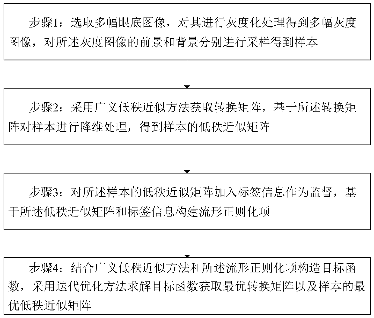
[0075] A classification model construction method for macular lesion region segmentation in fundus images, such as figure 1 shown, including the following steps:

[0076] Step 1: selecting multiple fundus images, performing grayscale processing on them to obtain multiple grayscale images, and sampling the foreground and background of the grayscale images respectively to obtain samples;

[0077] Step 2: Obtain the transformation matrix by using the generalized low-rank approximation method, and perform dimensionality reduction processing on the sample based on the transformation matrix to obtain the low-rank approximate matrix of the sample;

[0078] Step 3: adding label information to the low-rank approximate matrix of the sample as supervision, and constructing a regularization term based on the low-rank approximate matrix and label information;

[0079] Step 4: Combining the generalized low-rank approximation method and the regularization term to construct an objective func...

Embodiment 2

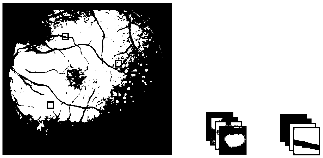
[0119] Based on the classification model in the first embodiment, this embodiment provides a method for segmenting the macular lesion region of the fundus image, which adopts the classification model in the first embodiment, including:

[0120] Step 1: Classify the test image based on the classification model to obtain the foreground point and the background point of the test image;

[0121] Step 2: Take the area where the foreground point is located as the segmentation result.

[0122] Among them, step 1 specifically includes:

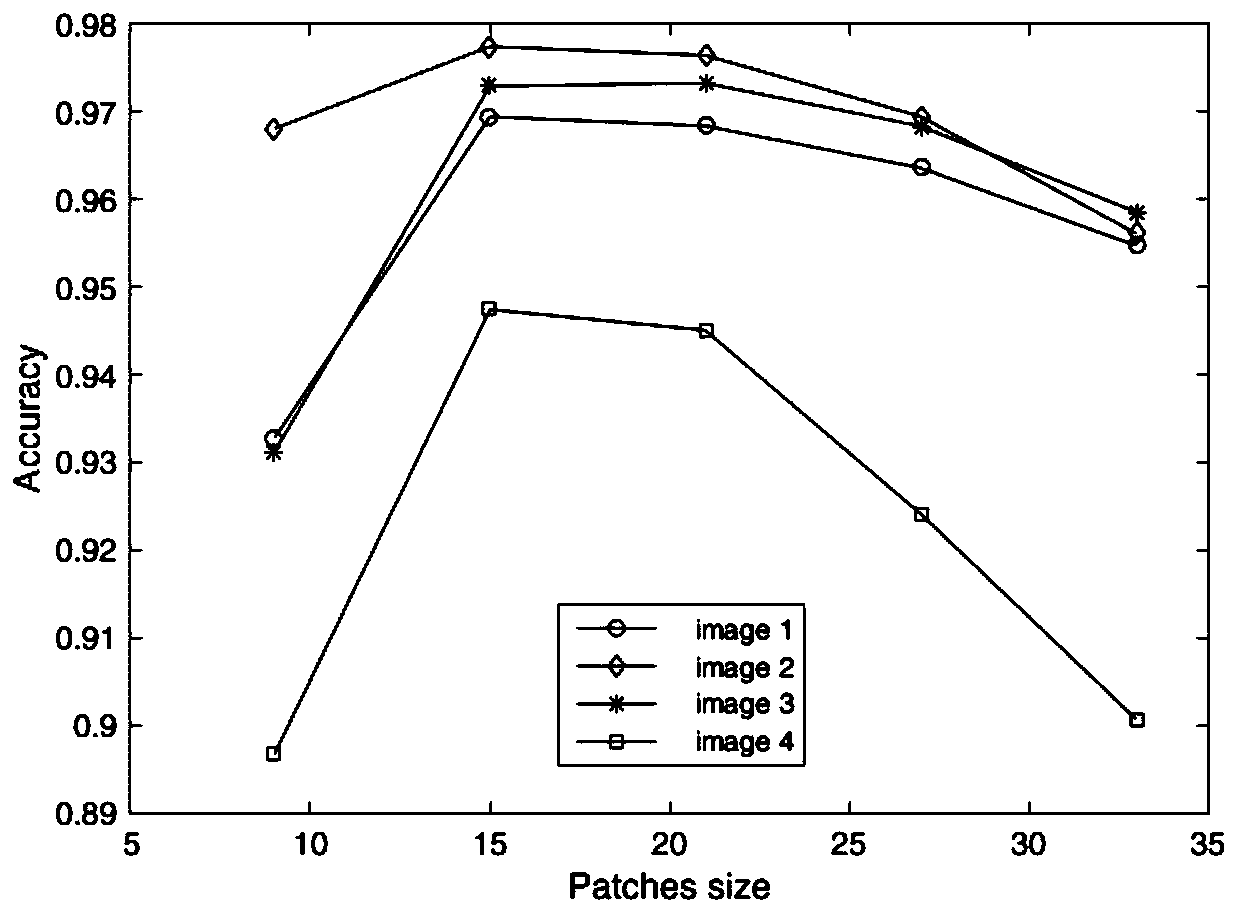
[0123] Grayscale the test image, scan the entire image with a k×k sliding window for sampling;

[0124] Using the optimal transformation matrix to reduce the dimensionality of the sample of the test image to obtain the optimal low-rank approximation matrix of the test image;

[0125] The optimal low-rank approximation matrix of the test image is used as the input of the SVM classifier to obtain the classification result.

[0126] If the test sample...

Embodiment 3

[0128] Based on the above image segmentation method, this embodiment provides a computer device for building a classification model for the segmentation of macular lesion regions in fundus images, including: a memory, a processor, and a computer program stored in the memory and operable on the processor , characterized in that, the processor implements the following steps when executing the program:

[0129] Receiving the user's selection of the fundus training image, performing grayscale processing on the training image to obtain a grayscale image; sampling the foreground and background of the grayscale image to obtain samples;

[0130] Obtaining a transformation matrix by using a generalized low-rank approximation method, performing dimensionality reduction processing on the sample based on the transformation matrix, and obtaining a low-rank approximation matrix of the sample;

[0131] Adding label information to the low-rank approximate matrix of the sample as supervision, ...


PUM


Abstract

The invention discloses a method for constructing a classification model for segmenting macular lesion regions of fundus images, which comprises the following steps: selecting multiple fundus images, performing grayscale processing on them to obtain multiple grayscale images, and performing grayscale processing on the grayscale images Samples are obtained by sampling the foreground and background of the sample respectively; the transformation matrix is ​​obtained by using the generalized low-rank approximation method, and the dimensionality reduction process is performed on the sample based on the transformation matrix to obtain the low-rank approximation matrix of the sample; the low-rank approximation matrix of the sample is added to The label information is used as supervision to construct the manifold regularization item; the objective function is constructed by combining the generalized low-rank approximation method and the manifold regularization item, and the iterative optimization method is used to solve the objective function to obtain the optimal transformation matrix and the optimal low-rank approximation of the sample matrix; constructing a classification model based on the optimal low-rank approximate matrix and label information. The classification model of the present invention can extract feature descriptors that are both low-dimensional and highly distinguishable, and can improve segmentation accuracy.

Description

technical field [0001] The invention relates to the field of medical image processing, in particular to a classification model construction method, device and image segmentation method for macular degeneration region segmentation in fundus images. Background technique [0002] Eyes are the most important organ for human beings to obtain information. The macula is located at the back of the eyeball and is an important tissue for people to perceive external light and objects. If the lesion occurs in this part, it will cause vision loss or even blindness, which is one of the important causes of blindness in the elderly. When doctors diagnose the macular degeneration region (drusen) in fundus images, there are disadvantages such as low accuracy, poor repeatability, and many subjective factors. Therefore, there is an urgent need for the application and research of macular degeneration region segmentation technology to meet the clinical needs of auxiliary medical care such as sc...

Claims


Application Information

Patent Timeline
Patent Type & Authority Patents(China)
IPC IPC(8): G06T7/11G06T7/194G06K9/62
CPCG06T7/11G06T7/194G06T2207/30096G06T2207/30041G06T2207/20081G06F18/2135G06F18/214G06F18/2411
Inventor 郑元杰任秀秀连剑刘弘赵艳娜秦茂玲
Owner SHANDONG NORMAL UNIV
Why Eureka
Try Eureka