Eureka AIR delivers breakthrough ideas for toughest innovation challenges, trusted by R&D personnel around the world.

Method for improving imaging stability of diasonograph 4D mechanical probe

An ultrasonic diagnostic instrument and technology of stability, applied in ultrasonic/sonic/infrasonic diagnosis, sonic diagnosis, infrasonic diagnosis and other directions, can solve the problems of 3D reconstruction image shaking and other problems, and achieve the effect of improving user experience and achieving remarkable results.

Active Publication Date: 2018-03-16
深圳纯和医药有限公司
View PDF7 Cites 2 Cited by
  • Summary
  • Abstract
  • Description
  • Claims
  • Application Information

AI Technical Summary

Problems solved by technology

[0006] The invention provides a method for improving the imaging stability of a 4D mechanical scanning probe of an ultrasonic diagnostic instrument, so as to solve the problem that the three-dimensional reconstruction image shakes with the vibration of the transducer caused by the forward and reverse gap of the mechanical transmission device

Method used


Image

  • Method for improving imaging stability of diasonograph 4D mechanical probe
  • Method for improving imaging stability of diasonograph 4D mechanical probe
  • Method for improving imaging stability of diasonograph 4D mechanical probe

Examples


Embodiment Construction

[0029] The embodiments of the present invention will be described in detail below with reference to the accompanying drawings, but the present invention can be implemented in many different ways defined and covered by the claims.

[0030] (1) Timing of two-dimensional tomographic image scanning

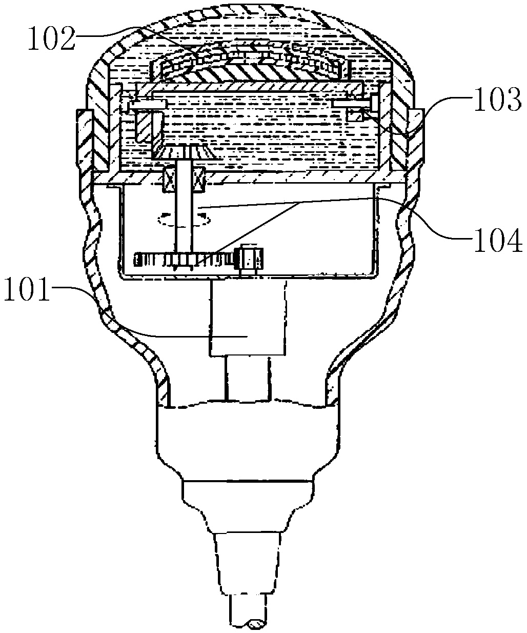
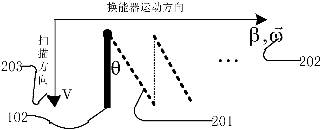
[0031] During 4D imaging, the transducer moves along figure 2 The indicated scanning direction continuously collects scanning lines, and a 2D tomographic image 201 is obtained in one scanning period. figure 2 indicated by the thick dashed line. Driven by the motor 101 , the transducer 102 rotates or translates at a speed 202 (the present invention is applicable to translation and rotation, and for simplicity of description, rotation is used as an example below). Due to the rotation of the transducer, the 2D tomographic image plane in space forms an angle θ with the transducer, figure 2 As shown, θ=ω*T, where ω is the moving speed of the transducer 202, and T is the scanning time...


PUM


Abstract

The invention provides a method for improving the imaging stability of a diasonograph 4D mechanical probe. The method comprises the steps of obtaining corresponding incomplete faultage image scanningduration in the process that a probe is rotated from the first position to the second position; obtaining transmission interval delay of a driving device for driving an energy converter to move in theprocess that the energy converter is rotated from the first position to the second position; in the process that the energy converter is rotated from the first position to the second position, afterthe energy converter driving device sends out an enabled signal for enabling the energy converter to start to move, after the first delay, starting to record and scan faultage image data through an imaging system till the energy converter is moved to the second position, wherein the first delay is introduced by the compensation transmission interval. The problem that three-dimensional images are twisted along with swinging of the energy converter due to the scanning time sequence and the transmission interval of a 4D mechanical scanning probe is solved.

Description

technical field [0001] The invention relates to the technical field of ultrasonic detection, in particular to a method for improving the three-dimensional imaging stability of a 4D mechanical scanning probe of a medical ultrasonic diagnostic instrument. Background technique [0002] Please refer to figure 1 The imaging process of the 4D mechanical scanning probe of the medical ultrasonic diagnostic instrument is as follows: the motor 101 drives the transducer array 102 (usually referred to as the transducer) to rotate or translate. The device collects spatial line data (or scanning lines), forms the line data into a two-dimensional tomographic image, that is, a 2D image, and then performs three-dimensional reconstruction on a group of 2D tomographic images corresponding to the spatial position, and displays the obtained three-dimensional image on the monitor. The above processes are executed sequentially or in parallel in real time to obtain real-time three-dimensional ultr...

Claims


Application Information

Patent Timeline
Patent Type & Authority Applications(China)
IPC IPC(8): A61B8/00
CPCA61B8/4444A61B8/4483A61B8/52
Inventor 陈颖川
Owner 深圳纯和医药有限公司
Why Eureka
Eureka Blog
Learn More