Segmentation method for inner membrane in blood vessel of intravascular unltrasound image

An ultrasound image and ultrasound image technology, applied in the field of image processing, can solve the problems of low robustness, inability to meet clinical real-time performance, redundant and complex model processing process, etc., and achieve the effect of preventing gradient disappearance and improving recognition ability.

Active Publication Date: 2018-04-13
SOUTH CHINA UNIV OF TECH
View PDF5 Cites 24 Cited by
  • Summary
  • Abstract
  • Description
  • Claims
  • Application Information

AI Technical Summary

Problems solved by technology

This type of method is highly dependent on the setting of the initial contour. Although there are corresponding processing methods designed for complex image features such as artifacts, plaques, and stents in intravascular ultrasound images, the processing process of the model is redundant and complicated. The robustness is low on high-frequency and high-resolution intravascular ultrasound images, which cannot meet the clinical real-time requirements

Method used


Image

  • Segmentation method for inner membrane in blood vessel of intravascular unltrasound image
  • Segmentation method for inner membrane in blood vessel of intravascular unltrasound image
  • Segmentation method for inner membrane in blood vessel of intravascular unltrasound image

Examples


Embodiment Construction

[0040] The present invention will be further described below in conjunction with specific examples.

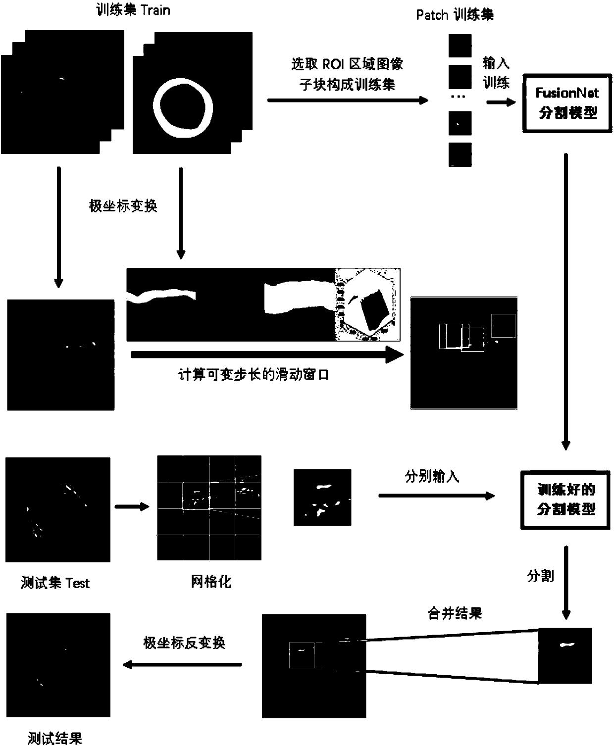
[0041] Such as figure 1 As shown, the method for intima segmentation of intravascular ultrasound images provided in this embodiment includes the following steps:

[0042] S1. Collect intravascular ultrasound image data, and mark the intima-media region in each intravascular ultrasound image.

[0043] Assume that a total of IVUS images I i , i∈[1,…,M], where M is the total number of images, for each I i Let the clinician label the medial area and there will be a corresponding labeling map L i , select I i and L i Four-fifths of them constitute the training set Train, and one-fifth constitute the independent test set Test. A total of 666 intravascular ultrasound images and annotations were collected, 500 of which were used as the training set and 166 were used as the test set Test. The image size is 512×512.

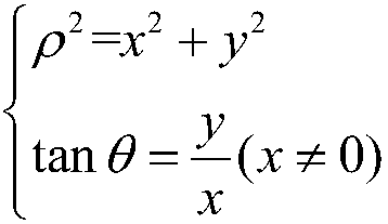
[0044] S2. Perform polar coordinate transformation on the tra...


PUM


Abstract

The invention discloses a segmentation method for an inner membrane in a blood vessel of an intravascular unltrasound image. The method comprises the steps of S1, collecting intravascular unltrasoundimage data, and marking a central inner membrane area in each intravascular unltrasound image; S2, performing polar coordinate transformation on a training set image; S3, computing an edge distance map of a mark chart; S4, setting a sliding clamping window and determining the size of the window and a sliding step length; S5, building and training a FusionNet deep learning segmentation model; and S6, inputting new data and acquiring a segmentation result. The method provided by the invention, the inner membrane area in the blood vessel can be quickly, accurately and effectively extracted.

Description

technical field [0001] The present invention relates to the technical field of image processing, in particular to a deep learning-based method for intima segmentation of intravascular ultrasound images. Background technique [0002] At present, cardiovascular disease has become one of the most important factors threatening human health. Among them, coronary atherosclerosis is an important cause of such diseases. If atherosclerosis can be identified and diagnosed in the early diagnosis, it will be of great significance to the diagnosis and treatment of coronary artery diseases. Intravascular Ultrasound (IVUS), which is currently used clinically, is an ultrasonic diagnostic method that can display the morphology of vessel walls and vascular plaques in real time. [0003] At present, for IVUS images collected clinically, doctors mainly focus on the region of interest (ROI) in the intima region of the blood vessel. In the actual diagnosis process, doctors often need to manually...

Claims


Application Information

Patent Timeline
Patent Type & Authority Applications(China)
IPC IPC(8): G06T7/11G06T7/12G06T7/194
CPCG06T7/11G06T7/12G06T7/194G06T2207/10132G06T2207/20021G06T2207/20081G06T2207/20084G06T2207/20104G06T2207/30101
Inventor 郭圣文吴宇鹏任力黄美萍梁稳生
Owner SOUTH CHINA UNIV OF TECH
Why Eureka
Try Eureka