Cell locating method and cell dividing method

A cell positioning and cell technology, applied in the field of biomedical image processing, can solve the problems of laborious and laborious, increase the difficulty of cell nucleus positioning cell segmentation, distinguish single cells, etc., and achieve the effect of improving accuracy

Active Publication Date: 2018-05-25
志诺维思(北京)基因科技有限公司
View PDF1 Cites 17 Cited by
  • Summary
  • Abstract
  • Description
  • Claims
  • Application Information

AI Technical Summary

Problems solved by technology

However, due to the adhesion and overlap between cells, similar phenomena are prone to occur in cell nucleus staining in cell staining images, making it difficult to quickly and accurately obtain cell nucleus position information and identify individual cells based on cell nucleus staining information, requiring professionals to specialize in staining images Carrying out cell nucleus information labeling and cell segmentation is labor-intensive
[0004] In addition, in the immunohistochemical staining images, the staining results of some molecular markers overlap with the staining results of cell nuclei, further increasing the difficulty of nuclei localization and cell segmentation

Method used


Image

  • Cell locating method and cell dividing method
  • Cell locating method and cell dividing method
  • Cell locating method and cell dividing method

Examples


Embodiment 1

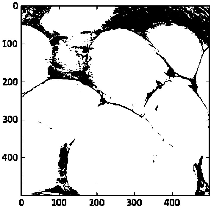
[0069] Such as figure 1 As shown, Embodiment 1 of the present invention provides a cell location method. The first stained image involved in the method is an HE stained colon cancer image, and the second stained image is an immunohistochemical stained image stained with a CD3 marker. The method specifically includes:

[0070] Step S1. Obtain the first stained image ( figure 1 The HE image shown, the source tissue is colon cancer tissue, the data set contains 100 500*500 images), the image includes nuclear staining (ie auxiliary staining) and artificially annotated nuclear location information (ie cell location information) .

[0071] Step S2. Take the first stained image as the learning object, and establish a prediction model for predicting the location of the cell nucleus based on the staining information of the cell nucleus through machine learning:

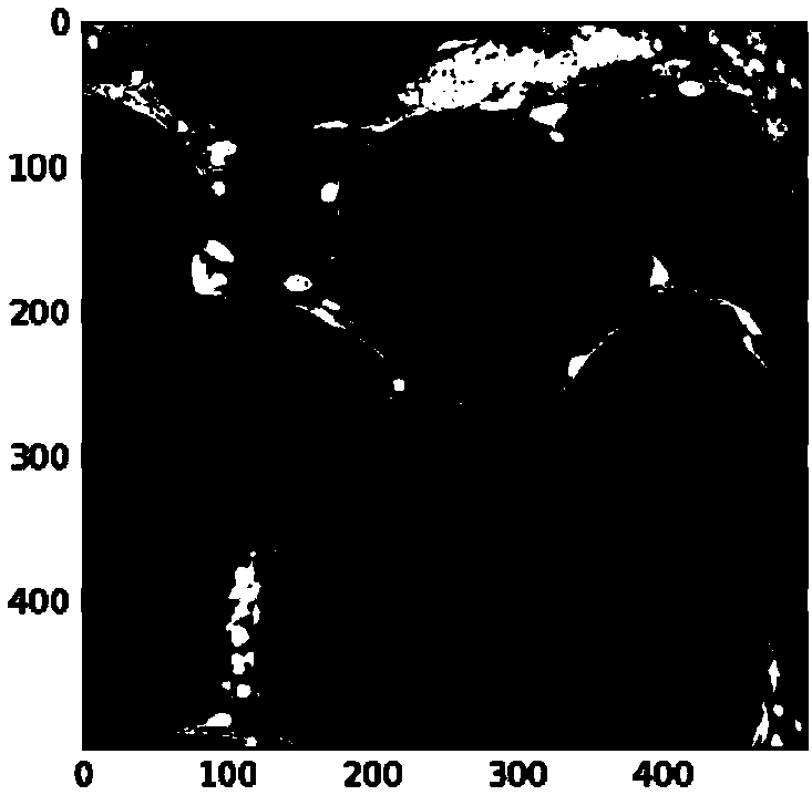
[0072] Step S201. Obtain the nuclear staining information of the first stained image (such as figure 2 Shown) and nuclear locati...


PUM


Abstract

The invention relates to a cell locating method and a cell dividing method. The cell locating method and the cell dividing method comprise the steps of conducting machine leaning on a first chromosomeimage containing cell locating information to obtain a prediction model, wherein the prediction model is used for a second chromosome image not containing the cell locating information and is used for predicting the cell locating information of the second chromosome image, and according to the cell locating information, conducting cell dividing on the second chromosome image. The cell locating method and the cell dividing method have the advantages of conducting automatic recognition in cell locating and cell dividing, not needing or reducing artificial identifications and saving time and labor.

Description

Technical field [0001] The present invention relates to the field of biomedical image processing, in particular to a method for cell positioning and a method for cell segmentation. Background technique [0002] Cell staining is a technical method widely used in clinical work and scientific research. The staining information reflects the cell morphology of the sample to be tested and the expression of molecular markers, which can provide clinicians and scientific researchers with important decision-making information. Among them, cell location and cell segmentation are of great significance in cell classification, counting, and analysis of staining results. [0003] Currently, cell location and cell segmentation are generally obtained through nuclear staining. However, due to the many adhesions and overlaps between cells, similar phenomena are likely to occur in cell staining images, which makes it difficult to quickly and accurately obtain nuclear position information and identify...

Claims


Application Information

Patent Timeline
Patent Type & Authority Applications(China)
IPC IPC(8): G06T7/00G06T7/11G06T7/136
CPCG06T7/0012G06T7/11G06T7/136G06T2207/20084
Inventor 刘小晴刘昌灵郝伶童凌少平
Owner 志诺维思(北京)基因科技有限公司
Why Eureka
Try Eureka