Infant brain magnetic resonance image partitioning method based on fully convolutional network

A magnetic resonance image and convolutional network technology, applied in image analysis, image enhancement, image data processing, etc.

Inactive Publication Date: 2018-06-15
SHENZHEN WEITESHI TECH
View PDF3 Cites 32 Cited by
  • Summary
  • Abstract
  • Description
  • Claims
  • Application Information

AI Technical Summary

Problems solved by technology

[0004] Aiming at the problem that gray matter and white matter have almost the same intensity level, the purpose of the present invention is to provide a method for brain MRI segmentation of infants and young children based on a fully convolutional network. Probability maps of the tissues, and then an initial segmentation of different brain tissues is obtained from the probability maps, which are used to calculate distance maps for each brain tissue, followed by modeling spatial context information from the distance maps, and finally by using spatial correlation information and multimodal magnetic resonance images To achieve the final segmentation, the training process mainly includes increasing training data, preparing training patches and iterative training, and evaluating with various detection values ​​after testing

Method used


Image

  • Infant brain magnetic resonance image partitioning method based on fully convolutional network
  • Infant brain magnetic resonance image partitioning method based on fully convolutional network
  • Infant brain magnetic resonance image partitioning method based on fully convolutional network

Examples


Embodiment Construction

[0034]It should be noted that, in the case of no conflict, the embodiments in the present application and the features in the embodiments can be combined with each other. The present invention will be further described in detail below in conjunction with the drawings and specific embodiments.

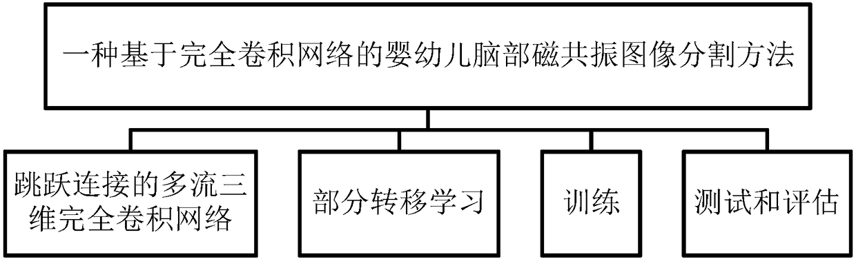
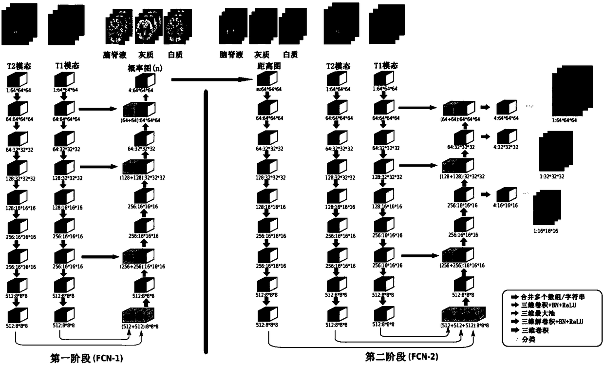
[0035] figure 1 It is a system framework diagram of a method for segmenting infant brain magnetic resonance images based on a fully convolutional network in the present invention. It mainly includes multi-stream 3D fully convolutional network (FCN) with skip connections, partial transfer learning, training, testing and evaluation.

[0036] In partial transfer learning, the weights of shallow layers in deep neural networks are general, while the weights of deep layers are more task-specific; in order to better take advantage of transfer learning, it is necessary to transfer from a training model of a related task; use A pre-trained model designed to segment the proximal femur from 3D T1...


PUM


Abstract

The invention provides an infant brain magnetic resonance image partitioning method based on a fully convolutional network. The main content of the method comprises the multi-stream three-dimensionalfully convolutional network (FCN) with jump connection, partial transfer learning, training and testing and evaluation. According to the process of the method, first, a probability graph of each pieceof brain tissue is learned from a multi-modal magnetic resonance image; second, initial partitions of different brain tissue are obtained from the probability graphs and used for calculating a distance graph of each piece of brain tissue; third, spatial context information is simulated according to the distance graphs; and last, final partitioning is realized by use of spatial correlation information and the multi-modal magnetic resonance image, wherein the training process mainly comprises training data increasing, training patch preparation and iterative training, and various detection values are used for evaluation after testing is performed. Through the partitioning method, a white matter area, a grey matter area and a cerebrospinal fluid area are successfully divided, the potential gradient vanishing problem of multi-level deep supervision is relieved, training efficiency is improved, and partitioning performance is greatly enhanced.

Description

technical field [0001] The invention relates to the field of image segmentation, in particular to a method for segmenting infant brain magnetic resonance images based on a fully convolutional network. Background technique [0002] Research on region of interest segmentation of medical images is the most important basis in medical image analysis. Accurate, robust and fast image segmentation is the most important step before quantitative analysis, 3D visualization and other follow-up links. Important clinical applications such as radiotherapy planning and treatment evaluation have laid the most fundamental foundation. Using automatic, accurate and quantitative computer-aided image analysis can help clinicians and researchers process massive image information efficiently and accurately. Brain segmentation in magnetic resonance (MR) images is a central part of this quantitative analysis tool, as it provides quantitative measurements of different brain structures and provides ba...

Claims


Application Information

Patent Timeline
Patent Type & Authority Applications(China)
IPC IPC(8): G06T7/10
CPCG06T7/10G06T2207/10088G06T2207/20081G06T2207/20084G06T2207/30016
Inventor 夏春秋
Owner SHENZHEN WEITESHI TECH
Why Eureka
Try Eureka