
A thin slice of simulated biological tissue, its preparation method and its application and device

A biological tissue and thin section technology, applied in the direction of measuring devices, test sample preparation, instruments, etc., can solve problems such as difficulty in cutting, difficulty in simulating the interaction between target molecules and actual tissues, difficulty in consistent sample slice shape and area, etc.

Active Publication Date: 2021-04-13
INST OF MATERIA MEDICA CHINESE ACAD OF MEDICAL SCI
View PDF7 Cites 0 Cited by
  • Summary
  • Abstract
  • Description
  • Claims
  • Application Information

AI Technical Summary

Problems solved by technology

However, the above-mentioned preparation schemes all have deficiencies: using the homogenate slice method to prepare simulated tissue, the texture of the frozen block of homogenate tissue is soft and brittle, and it is not easy to cut, and the shape and area of ​​the slices between samples are difficult to be consistent, and the tissue homogenization During the process, the cell damage rate is extremely high, and it is difficult to simulate the interaction between the target molecule and the actual tissue; the method of using a micropipette to drop the target molecule on the surface of the tissue is not suitable for small-sized tissue samples, and the target molecule Which organ level and tissue micro-area to drop to is completely selected by the operator, and the specific matrix effect of different tissue areas has different effects on the target molecules, so it is difficult to guarantee the parallelism of the results between different batches of samples; Although the spraying method can ensure the uniform coating of target molecules, since the target molecules are distributed on the surface of biological tissue slices and do not fully penetrate deep into the tissue, the influence of tissue matrix effect on the signal response intensity is significantly lower than that of actual biological samples. The effect of the target molecule

Method used


Image

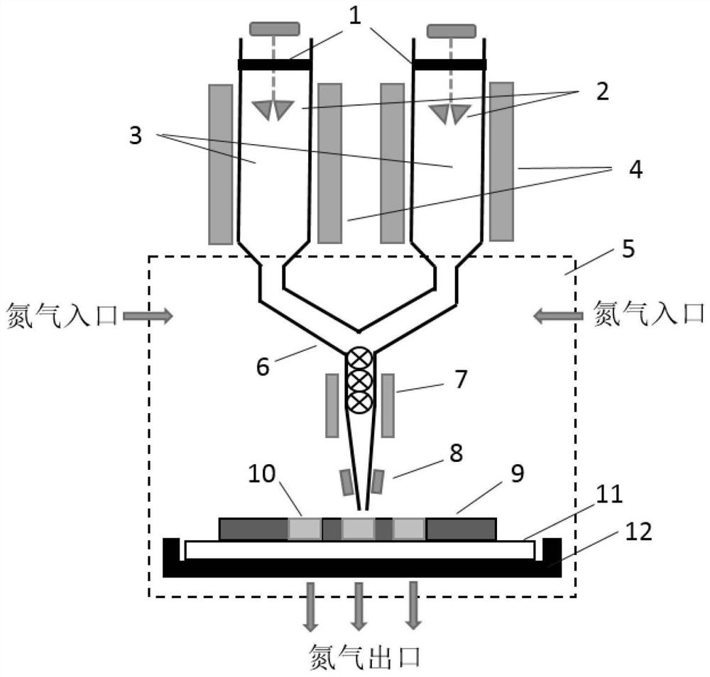
  • A thin slice of simulated biological tissue, its preparation method and its application and device
  • A thin slice of simulated biological tissue, its preparation method and its application and device
  • A thin slice of simulated biological tissue, its preparation method and its application and device

Examples


Embodiment 1

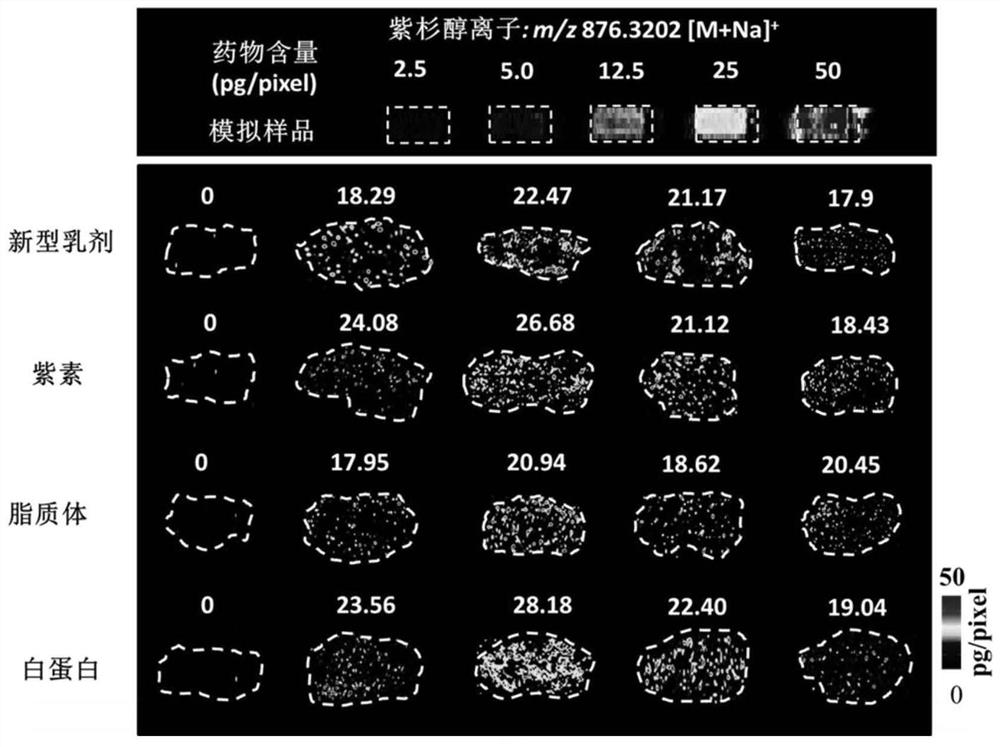
[0154] Example 1: Quantitative visualization study on the distribution of paclitaxel in tumor tissue

[0155] (1) Sample preparation

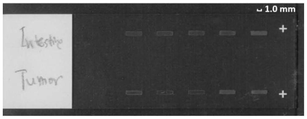
[0156] Polyvinyl chloride (PVC) film was used as the shaped material for simulating biological tissue slices, and five rectangular shaped template frames (4mm×1mm) were engraved continuously in the same row by a high-precision CNC mechanical cutting plotter. Before use, tear off the protective film on the back of the engraved PVC template frame, add a small amount of ultra-pure water to soak the sticky side, dry it, and then paste it on the positive charge anti-off glass slide to form a shape together with the slide rectangular mold slots.

[0157] Paclitaxel standard solutions with different concentrations were added into ultrapure water as the aqueous dispersion medium.

[0158] Weigh 400 mg of xenograft tumor tissue from Balb / C female nude mice, cut it into pieces, add 1.0 mL of aqueous dispersion medium containing paclitaxel standard solu...

Embodiment 2

[0169] Example 2: Rapid quantitative screening of several potential markers of esophageal cancer in esophageal tissue

[0170] (1) Sample preparation

[0171] Polyvinyl chloride (PVC) film is used as the forming film material for simulating biological tissue slices, and the high-precision numerical control mechanical cutting plotter is continuously engraving 4 lines, and each line has 5 rectangular shaped template frames (4mm×1mm). Before use, tear off the protective film on the back of the engraved PVC template frame, add a small amount of ultra-pure water to soak the sticky side, dry it, and then paste it on the positive charge anti-off glass slide to form a shape together with the slide rectangular mold slots.

[0172] Stable isotope labeled mixed standard solution (L-arginine- 13 C 6 , L-carnitine-d 3 and phosphatidylcholine(18:0)-d 13 ) into ultrapure water as an aqueous dispersion medium.

[0173] Weigh the control esophageal tissue sample, cut it into pieces, add ...

Embodiment 3

[0181] Example 3: Similarity Evaluation and Preparation Method Verification of Simulated Biological Tissue Thin Sections

[0182] The simulated tissue thin slices prepared in Example 1, together with the real tumor tissue section (8 μm), were subjected to hematoxylin-eosin staining (H&E staining), and qualitatively evaluated the similarity between the simulated tissue and the real tissue from the aspect of histomorphology (results see attached Figure 7 ).

[0183] Take 5 copies of simulated biological tissue slices and actual biological tissue slices, and electronic balance (1 / 100,000) accurately weighs the quality difference of the glass slides before and after adding samples, and calculates the quality of each simulated tissue slice and actual tissue section; collect the above For the optical images of 10 slices, use the Photoshop image processing software to sum the number of pixels in the sample area, convert the pixel size and the scale into the actual area of ​​each sa...


PUM

PropertyMeasurementUnit
angleaaaaaaaaaa

Abstract

The invention belongs to the technical field of analysis and detection, and relates to a simulated biological tissue slice, a preparation method thereof, an application and a device thereof. The simulated biological tissue slice has regular shape and controllable size, mainly consists of biological tissue material and tissue cells, and contains a known amount of target molecules, which can ideally simulate the matrix of real biological tissue slices. The preparation method is quick and easy, and has high repeatability. The simulated biological tissue standard slice preparation device can realize the automatic preparation of simulated biological tissue. The simulated biological tissue thin slice prepared by the invention provides a stable and controllable standard biological tissue sample for quantitative mass spectrometry imaging analysis and mass spectrometry high-throughput rapid detection of target molecules such as drugs and diagnostic markers. It has important application value in the fields of preclinical drug development and clinical disease diagnosis.

Description

technical field [0001] The invention belongs to the technical field of biological analysis and detection, and relates to a thin slice of simulated biological tissue, its preparation method and its application and device, which are applied to quantitative mass spectrometry imaging analysis and rapid mass spectrometry detection. [0002] technical background [0003] Mass spectrometry imaging technology is based on mass spectrometry analysis. Various molecules on the surface of the sample to be tested (mostly biological tissues) are detected according to their spatial positions, and the relationship between their mass-to-charge ratio intensity and position is obtained to form multidimensional data. Through data processing and software Reconstruct the image visualization of ion intensity, and finally realize the molecular imaging technology of simultaneous detection of multiple molecules and display their spatial distribution. It does not require radioactive isotopes, fluorescen...

Claims


Application Information

Patent Timeline
Patent Type & Authority Patents(China)
IPC IPC(8): G01N1/28G01N27/62
CPCG01N1/28G01N27/62
Inventor 再帕尔阿不力孜宋肖炜贺玖明厉欣孙成龙罗志刚李铁钢黄罗娇
Owner INST OF MATERIA MEDICA CHINESE ACAD OF MEDICAL SCI
Learn More