Dual-source CT coronary artery automatic extraction method

A coronary artery, automatic extraction technology, applied in the field of medical image processing, can solve problems such as manual operation and branch detection difficulties, and achieve the effect of reducing uneven grayscale and noise

Active Publication Date: 2018-11-06
GUANGDONG PHARMA UNIV
View PDF2 Cites 11 Cited by
  • Summary
  • Abstract
  • Description
  • Claims
  • Application Information

AI Technical Summary

Problems solved by technology

[0018] The present invention aims at the problems of difficult branch detection and manual operation in the existing heart coronary artery segmentation method, and simultaneously considers the gray scale characteristics, morphological characteristics and spatial relationship of blood vessels, and combines the statistical classification method to improve the multi-scale filtering and spherical model, Realize automatic three-dimensional coronary artery segmentation without doctor's interactive point selection

Method used


Image

  • Dual-source CT coronary artery automatic extraction method
  • Dual-source CT coronary artery automatic extraction method
  • Dual-source CT coronary artery automatic extraction method

Examples


Embodiment 1

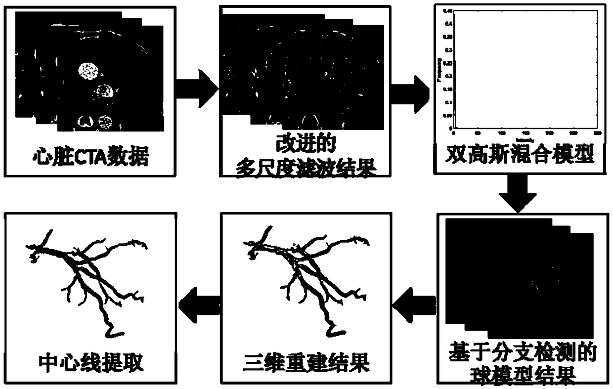
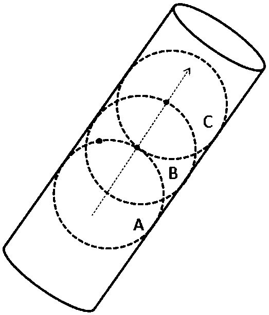
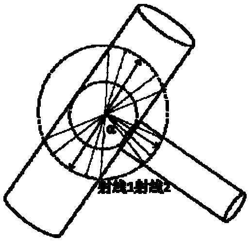
[0048] The present invention starts from the three aspects of blood vessel shape, gray feature and neighborhood relationship to segment coronary arteries simultaneously. The shape of the blood vessel is tubular. By calculating the eigenvalue of the Hessian matrix for each pixel, the Rb operator of the frangi model is improved, and a new blood vessel model is established to enhance the blood vessel structure. In the enhanced image, the gray scale of the blood vessel area is brighter, and the background area is darker. Using a statistical model, Gaussian modeling is performed on the blood vessel class and the background class. In the blood vessel class, the neighborhood relationship is constrained by the growth of the ball model, and coronary arteries are grown. To avoid false branch vessels and save time, a branch detection based on hierarchical clustering is established. The overall flowchart of the specific implementation method of the present invention is as figure 1 shown...


PUM


Abstract

The invention relates to a dual-source CT coronary artery automatic extraction method. The method comprises the following steps of S1, calculating eigenvalues lambda 1, lambda 2 and lambda 3 of a Hessian matrix for each pixel point in dual-source CT data, and building a new blood vessel model equation; effectively enhancing a blood vessel structure by utilizing the new blood vessel model; enablinga blood vessel region to be relatively bright in grayscale, and enabling a background region to be relatively dark; performing Gaussian modeling on the blood vessel region and the background region by using a statistical model; S2, by utilizing a Gaussian mixture model, classifying enhanced data into two types, namely, the background region and the blood vessel region; and S3, in the blood vesselregion, restraining a neighborhood relation through the growth of a ball model, growing coronary arteries, and detecting and removing pseudo-branch blood vessels in the grown coronary arteries by utilizing hierarchical clustering-based branch detection.

Description

technical field [0001] The invention relates to the field of medical image processing, in particular to a dual-source CT coronary artery automatic extraction method. Background technique [0002] Coronary artery segmentation is the first step in image analysis for cardiovascular disease. CT angiography is one of the most common examination methods at present. The amount of CT angiography data is large, the individual differences of patients are large, and the cardiovascular structure is complex. At present, the coronary artery segmentation method requires manual interaction, and the problem of pipeline branch error is prone to occur during pipeline reconstruction, which directly affects the doctor's analysis of blood vessels. Therefore, most doctors are still using two-dimensional images such as Multi-planner reformation (MPR), Curved planner reformation (CPR) and maximum intensity projection (MIP) to observe blood vessels. The traditional method analyzes blood vessels to...

Claims


Application Information

Patent Timeline
Patent Type & Authority Applications(China)
IPC IPC(8): G06T7/00G06T7/11
CPCG06T7/0012G06T2207/10081G06T2207/30101G06T7/11
Inventor 赵洁蒋世忠黄展鹏
Owner GUANGDONG PHARMA UNIV
Why Eureka
Try Eureka