A method for locating the position of the first rupture of a thoracic aortic dissecting aneurysm

A technology for dissecting aneurysm and thoracic aorta, which is applied in image analysis, image enhancement, instruments, etc., can solve the problems of increasing the pain of patients, finding the position of the first breach, and the injection concentration of contrast agent should not be too high, and achieve the positioning time. Short, pain-relieving effect

Active Publication Date: 2022-08-02
SOUTHEAST UNIV
View PDF5 Cites 0 Cited by
  • Summary
  • Abstract
  • Description
  • Claims
  • Application Information

AI Technical Summary

Problems solved by technology

However, due to the limited frame rate of DSA images and the injection concentration of contrast agent should not be too high, it is difficult to directly find the first breach location based on DSA images
At the same time, in order to reduce breathing artifacts, the patient is usually in a state of respiratory arrest during the shooting process, which increases the pain of the patient

Method used


Image

  • A method for locating the position of the first rupture of a thoracic aortic dissecting aneurysm
  • A method for locating the position of the first rupture of a thoracic aortic dissecting aneurysm
  • A method for locating the position of the first rupture of a thoracic aortic dissecting aneurysm

Examples


Embodiment 1

[0051] The patent of the present invention will be described in further detail below in conjunction with the accompanying drawings and embodiments, figure 1 It is a general flow chart of the present invention. In this embodiment, the imaging equipment is: PHILIPS AlluraXper FD20; the contrast agent is: Omnipac (specification of iodine content: 350mg / ml, dosage 100ml), which is a product of (General Electric Pharmaceuticals (Shanghai) Co., Ltd.). The contrast agent contrast method was as follows: catheterization through the femoral artery, using a high-pressure syringe (Medrad Mark V Provis) to inject the contrast agent into the aorta through a 5F gold-marked pigtail catheter, the imaging speed was 6 frames / s, and the injection pressure was 600 psi (4136.85 psi). kPa). In this embodiment, the software used for the algorithm is Matlab 2010.

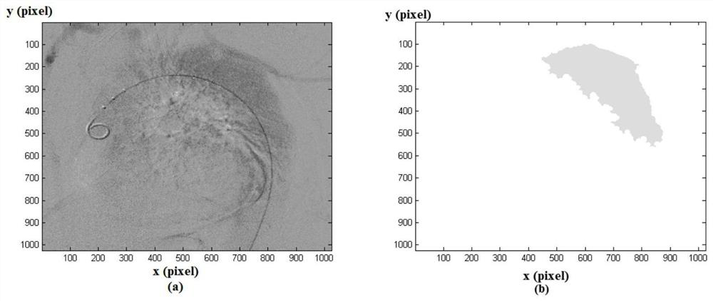
[0052] Step 1: Segment the aneurysm according to the difference in the fading time between the aneurysm and the blood vessel after the i...


PUM


Abstract

The invention relates to the technical field of post-processing of medical images, in particular to a method for locating the position of the first breach of a thoracic aortic dissection aneurysm based on digital subtraction angiography images. The aneurysm was segmented by the difference of the contrast agent; secondly, the cardiac cycle was segmented using the step-like change characteristics of the area of ​​the area in which the fluid was perfused by the contrast agent; thirdly, the first cardiac cycle of the hemangioma area after the contrast agent began to enter the area was obtained. The time-integrated image in the cycle is obtained, and its intensity distribution curve is obtained, and the true cavity and the false cavity are divided according to the intensity distribution curve; The first breach is located; the success rate of this method is about 85%. Compared with the manual positioning method, the method has a shorter positioning time, and the method can shorten the image acquisition time and reduce the injection volume of the contrast agent.

Description

technical field [0001] The invention relates to the technical field of post-processing of medical images, in particular to a method for locating the position of the first rupture of a thoracic aortic dissection aneurysm based on digital subtraction angiography images. Background technique [0002] Aortic dissecting aneurysm generally refers to the aortic intima torn due to various reasons, the high-pressure blood flow of the artery enters the aortic media through the tear, and the arterial media is separated from the adventitia to form a false lumen. Its pathological basis is the degeneration of the medial elastic fibers and smooth muscle of the aortic wall. Aortic dissecting aneurysm has an acute onset and a high mortality rate. Early and accurate diagnosis is the key to achieving significant therapeutic effects. [0003] The purpose of surgical treatment of aortic dissection is to select the surgical method according to the location of the dissection tear, the extent of t...

Claims


Application Information

Patent Timeline
Patent Type & Authority Patents(China)
IPC IPC(8): G06T7/00G06T7/11G06T7/187G06T7/62
CPCG06T7/0012G06T7/11G06T7/187G06T7/62G06T2207/30101G06T2207/10081
Inventor 黄朔朱斌孙钰周平万遂人程乐张冰
Owner SOUTHEAST UNIV
Why Eureka
Try Eureka