Method and magnetic resonance system to optimize mr images

a magnetic resonance and image technology, applied in image enhancement, instruments, applications, etc., can solve the problem of not finding the global optimum in the two-dimensional search region, and achieve the effect of simplifying non-contrast agent-enhanced mr angiography procedures, simple and fast manner

Inactive Publication Date: 2009-03-12
SIEMENS AG
View PDF7 Cites 32 Cited by
  • Summary
  • Abstract
  • Description
  • Claims
  • Application Information

AI Technical Summary

Benefits of technology

[0007]An object of the present invention is to simplify non-contrast agent-enhanced MR angiography procedures insofar so that the correct imaging parameters can be determined in a simpler and quicker manner.
[0008]This object is achieved in accordance with the invention by a method for optimization of angiographic magnetic resonance images in which veins and arteries can be presented separately, wherein multiple MR overview images are acquired, and at least one imaging parameter is varied in the acquisitions of the MR overview images. At least one optimized imaging parameter is subsequently automatically calculated using a quality criterion, and the optimized imaging parameter(s) is / are provided for the acquisition of the angiographic magnetic resonance images in which arteries can be shown separately from the veins. In the method according to the present invention, the operator no longer needs to study MR overview images in order to determine the imaging parameter(s) with which arteries and veins can be separated. The operator is thus unburdened, the personnel do not need to be specially trained for this angiography method, and the residence time of an examination subject in the magnetic resonance system is shortened since an automatic determination of an optimized imaging parameter is distinctly faster and less error-prone than a manual determination by means of viewing multiple MR images.
[0014]The use of the three-dimensional excitation volume of which the angiography exposures should be acquired to generate a two-dimensional overview image is an important step for the continuing automation of the method since an extra positioning step to position the excitation volume for the overview images is omitted. For excitation of a thinner slice, as is typically the case in a two-dimensional measurement, it would first have to be ensured by the operator that the vessel to be presented is contained at all in the excited volume. The use of the three-dimensional imaging sequence with deactivated phase coding in one direction furthermore has the advantage that the same sequence scheme (and therefore the same flow sensitivity as for the subsequent actual angiography measurement) is used for the determination of the quality criterion. A 2D turbo spin echo sequence switches gradients (typically a few) that are necessary in order to suppress an unwanted signal from imperfect refocusing pulses, different than a 3D turbo spin echo sequence. It therefore also has a different flow sensitivity.
[0016]According to a further embodiment of the invention, the MR overview images or the difference images can be masked or filtered. The goal of the masking or filtering is to avoid having to consider, or to consider to a lesser degree, pixels in overview images or difference images that are outside of a predetermined region. Dependent on the conoral orientation of the MR images, for example, the signal intensities at the upper and lower edges of the MR image in the direction of the body axis are typically subject to distortions. This is a consequence of the inhomogeneity of the B0 field in this region. These distortions can lead to errors in the determination of the quality criterion. This is prevented by the masking or filtering of these regions.
[0020]In an embodiment, it is possible to scan the cardiac cycle with a trigger delay change ΔTD in steps, such that the cardiac cycle is examined with different trigger delays that respectively differ by ΔTD within an R-spike (R-peak) interval. In another embodiment, a first optimization phase (run-through) is implemented in which the trigger delay TF is varied in larger steps, and from this first rough trigger delays TDSys and TDDia are calculated, while in a second optimization phase the trigger delays are varied in smaller steps and in a smaller search range in order to more precisely determine the trigger delays TDSys and TDDia determined in the first phase. Overall, the acquisition time to acquire the overview images can be shortened via the two-part optimization since overall fewer overview images must be acquired in comparison to the embodiment in which the cardiac cycle is examined in small trigger delay steps in one pass. In the prior art, a two-stage method would not lead to a reduction of the total examination duration, since the additional time that the operator requires to view the images after the first step and to determine the imaging parameters for the second step will generally be longer than the measurement time saved by the smaller total count of the overview images.

Problems solved by technology

This optimization in two steps is generally quicker than the exhaustive search in a two-dimensional search region, but generally does not find the global optimum in the two-dimensional search region.

Method used


Image

  • Method and magnetic resonance system to optimize mr images
  • Method and magnetic resonance system to optimize mr images
  • Method and magnetic resonance system to optimize mr images

Examples


Embodiment Construction

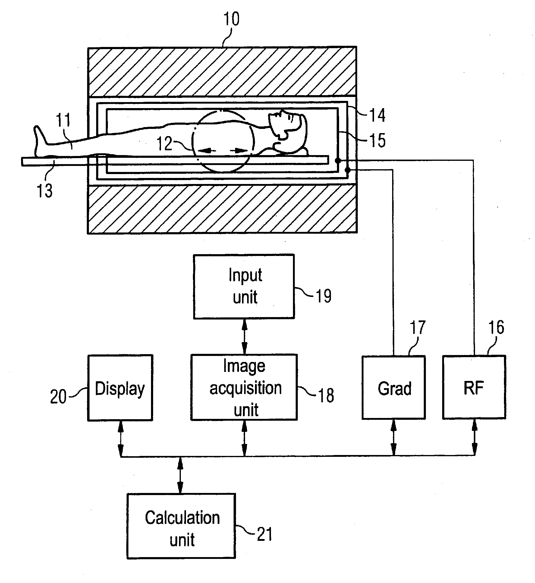
[0029]An MR system with which an imaging parameter can be optimized in a simple manner before conducting an angiographic measurement is schematically presented in FIG. 1. Such an MR system has a magnet 10 for generation of a polarization field B0. An examination subject (here an examination subject 11) is moved on a bed 13 into the magnet 10, as is schematically depicted by the arrows 12. The MR system furthermore has a gradient system 14 for generation of magnetic field gradients that are used for the imaging and spatial coding. To excite spins that are polarized due to the basic magnetic field, a radio-frequency coil arrangement 15 is provided that radiates a radio-frequency field into the examination subject 11 in order to deflect the magnetization from the equilibrium (steady) state. A gradient unit 17 is provided to control the magnetic field gradients and a RF unit 16 is provided to control the radiated RF pulses. An image acquisition unit 18 centrally controls the magnetic re...


PUM


Abstract

In a method and magnetic resonance MR system for the optimization of angiographic MR images of an examination subject, in which arteries can be presented separately from veins in the angiographic magnetic resonance images, multiple MR overview images are acquired, with at least one imaging parameter being varied in the acquisitions of the MR overview images, at least one optimized imaging parameter is automatically calculated using a quality criterion, and the optimized imaging parameter is provided for the acquisition of the angiographic magnetic resonance images in which arteries can be shown separately from the veins.

Description

BACKGROUND OF THE INVENTION[0001]1. Field of the Invention[0002]The present invention concerns a method to optimize angiographic magnetic resonance (MR) images of an examination subject and a magnetic resonance system that implements such a method. The invention is particularly applicable in the generation of peripheral MR angiographs in which the angiographic images are generated without using a contrast agent.[0003]2. Description of the Prior Art[0004]One possibility to generate magnetic resonance angiographs without the use of contrast agents is the employment of fast spin echo imaging sequences, wherein a three-dimensional turbo spin echo imaging sequence is combined with a technique known as the half Fourier technique, for example. In the half Fourier technique, one half of Fourier space (domain) or k-space is not completely filled with measurement data, and the data that are not acquired are calculated by symmetry requirements of the data. Given suitable parameterization of th...

Claims


Application Information

Patent Timeline
Patent Type & Authority Applications(United States)
IPC IPC(8): A61B5/055G06K9/00
CPCA61B5/0456A61B5/055A61B5/7285G01R33/5635G01R33/5673G06T2207/30101G06T7/0012G06T7/0081G06T7/0097G06T2207/10088G06T2207/20224G06T5/50A61B5/352G06T7/11G06T7/174
Inventor STEMMER, ALTO
Owner SIEMENS AG
Why Eureka
Try Eureka