Craniocerebral simulation model and preparation method thereof

A simulation model, brain technology, applied in the field of biomedicine, can solve problems such as simulation, and achieve the effect of avoiding errors

Active Publication Date: 2018-11-20
MEDPRIN REGENERATIVE MEDICAL TECH
View PDF10 Cites 7 Cited by
  • Summary
  • Abstract
  • Description
  • Claims
  • Application Information

AI Technical Summary

Problems solved by technology

[0005] The purpose of the present invention is to overcome the defect that the existing brain model cannot simulate

Method used


Image

  • Craniocerebral simulation model and preparation method thereof
  • Craniocerebral simulation model and preparation method thereof
  • Craniocerebral simulation model and preparation method thereof

Examples


Example Embodiment

[0062] Example 1

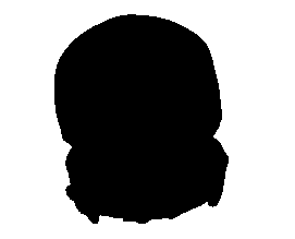
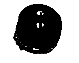
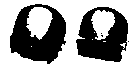
[0063] Such as Figure 1~10 As shown, a simulation model of the brain includes an upper skull, a lower skull and brain tissue, the upper skull and the lower skull are detachably fixedly connected to form a skull cavity, and the brain tissue is located in the skull cavity; the brain The tissue is prepared by the following process: the upper brain tissue mold is detachably sealed and fixedly connected with the lower skull bone provided with the lower brain tissue mold inside the cavity, and then the upper brain tissue is filled with silica gel or hydrogel material and cured; wherein, the upper brain tissue The mold and the lower brain tissue mold have contours and grooves that match the outer surfaces of the upper brain tissue and the lower brain tissue, respectively.

[0064] The upper brain tissue mold is connected with the lower skull through a slot or a vertical latch. The connection structure and connection method between the upper skull and the lower skull a...


PUM

PropertyMeasurementUnit
Thicknessaaaaaaaaaa
Thicknessaaaaaaaaaa

Abstract

The invention relates to a craniocerebral simulation model and a preparation method thereof. The model comprises an upper skull, a lower skull and a brain tissue, wherein the upper skull and the lowerskull are detachably fixedly connected and form a skull cavity, and the brain tissue is located in the skull cavity; The brain tissue is prepared through the following process: after an upper brain tissue mold is detachably fixedly connected with the lower skull with the lower brain tissue mold arranged in a cavity body in a sealed manner, the brain tissue is obtained by silica gel pouring or hydrogel material solidification; and the upper brain tissue mold and the lower brain tissue mold have contours and gullys matched with outer surfaces of an upper brain tissue and a lower brain tissue respectively. The brain tissue in the model provided by the invention has the flexibility, elasticity and touch similar to a real brain tissue; and is almost error-free in structure compared with the structure of the real brain tissue, and can be applied to the fields of medical teaching training, clinical operation planning, approach design, preoperative simulation, operation drill and the like.

Description

technical field [0001] The invention belongs to the field of biomedicine, and more specifically relates to a brain simulation model and a preparation method thereof. Background technique [0002] The models in the prior art that imitate the structure of the human brain can only roughly simulate the structure, the accuracy is very low, and the performance and tissue touch are very different from the real human brain structure, which is not suitable for medical anatomy. and clinical application. At present, clinical anatomy teaching mainly uses human brain specimens. However, human brain specimens are scarce and expensive, and because they need to be stored in formalin solution, the smell is unpleasant, which has a lot of inconvenience for the demonstration teaching of brain anatomy. Furthermore, the existing cranial brain models all use molds for standardized production, which cannot be individually prepared and cannot be used clinically. [0003] The 3D printed brain mode...

Claims


Application Information

Patent Timeline
IPC IPC(8): G09B23/34
CPCG09B23/34
Inventor 范小敏邓坤学袁玉宇
Owner MEDPRIN REGENERATIVE MEDICAL TECH
Why Eureka
Try Eureka