Construction method and application of thoracic disease detection model

A chest disease and detection model technology, applied in the field of medical imaging, can solve problems such as difficulty in ensuring diagnostic efficiency and accuracy, and achieve the effects of improving diagnostic efficiency and accuracy, reducing diagnostic time, and improving efficiency and accuracy

Active Publication Date: 2018-11-27
HUIYING MEDICAL TECH (BEIJING) CO LTD
View PDF3 Cites 25 Cited by
  • Summary
  • Abstract
  • Description
  • Claims
  • Application Information

AI Technical Summary

Problems solved by technology

However, due to the lack of a large number of professional medical imaging doctors, it is difficult to guarantee the efficiency and accuracy of diagnosis

Method used


Image

  • Construction method and application of thoracic disease detection model
  • Construction method and application of thoracic disease detection model
  • Construction method and application of thoracic disease detection model

Examples


Embodiment 1

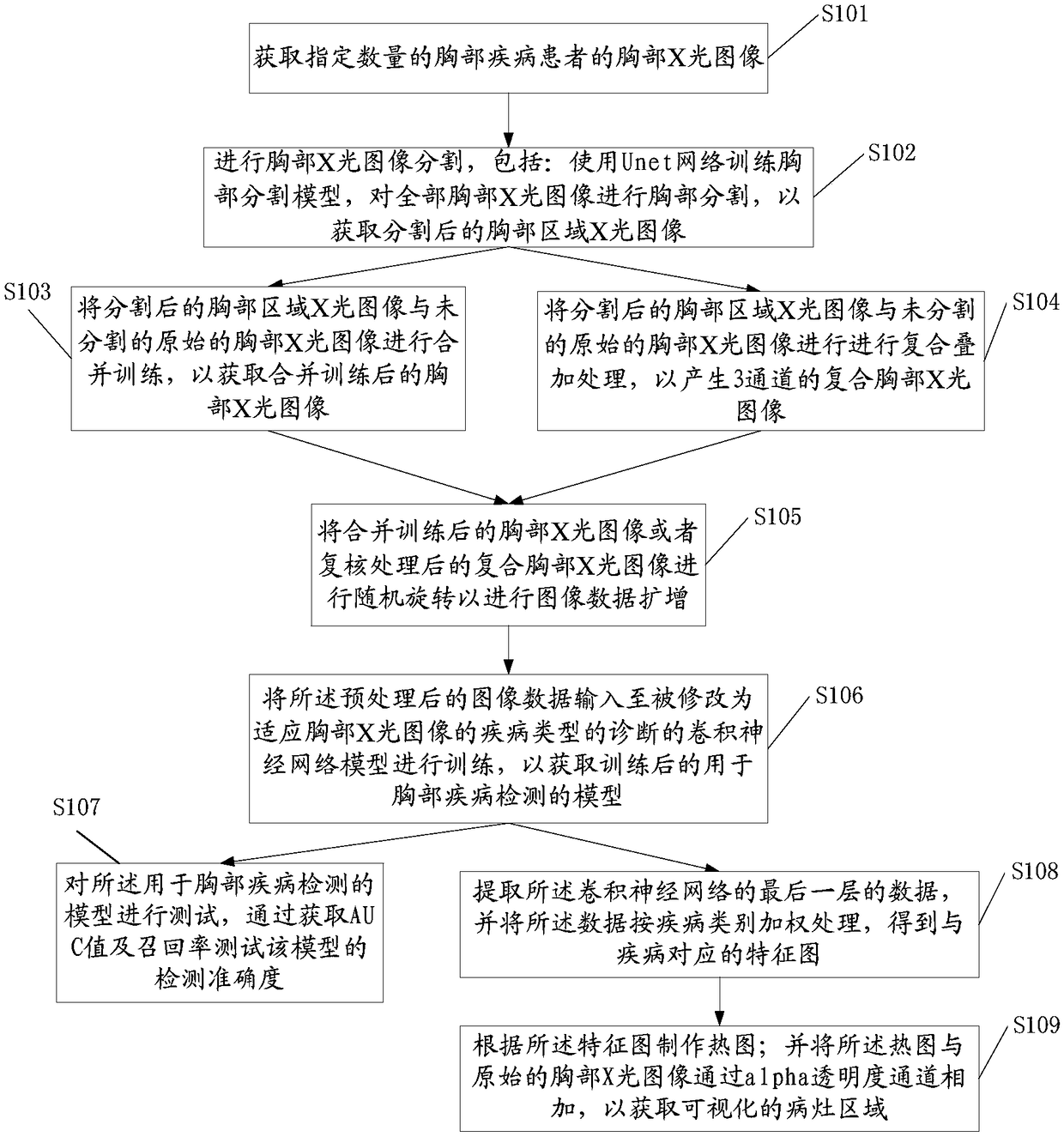
[0046] Such as figure 1 As shown, the application provides a method for constructing a chest disease detection model, including:

[0047] S101. Acquire chest X-ray images of a specified number of chest disease patients; the chest X-ray images may use a large number of existing chest X-ray images of chest disease patients.

[0048] Then in steps S102-104, image preprocessing is performed on the X-ray image to obtain preprocessed image data.

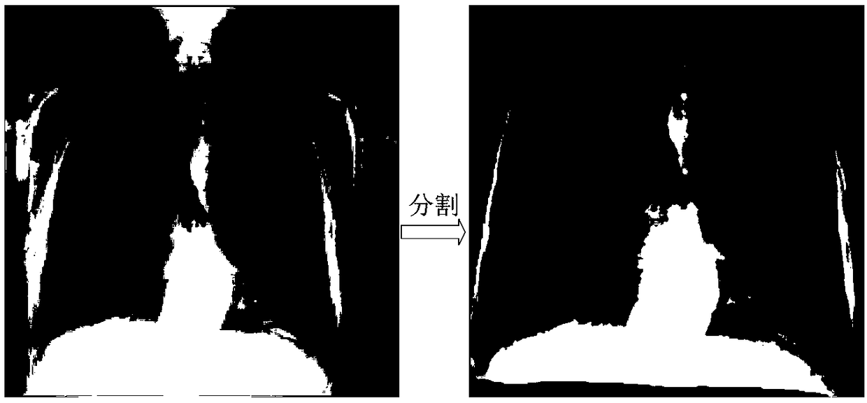
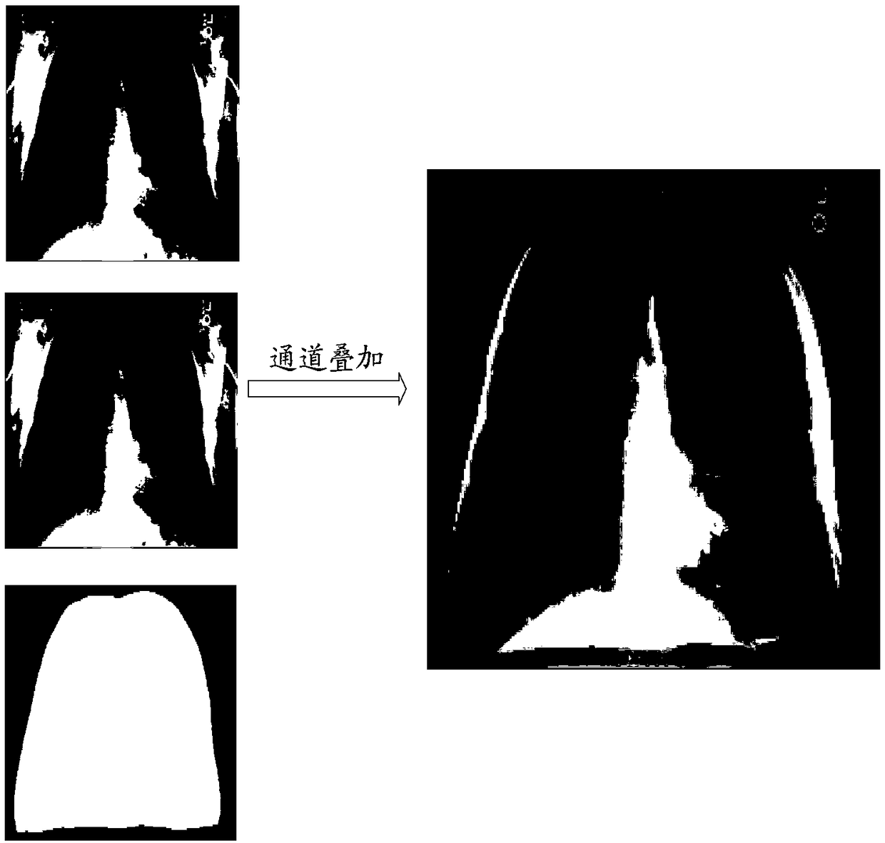
[0049] S102, performing chest X-ray image segmentation, including: using the Unet network to train a chest segmentation model, and performing chest segmentation on all chest X-ray images to obtain segmented chest X-ray images. Specifically, it includes: B11. Preliminarily locate the chest area through connected domain analysis. B12. Delete the non-image area at the edge of the chest area. Such as figure 2 As shown, the picture on the right shows that the segmented chest radiograph (chest X-ray image) can enable the Unet network to acc...

Embodiment 2

[0071] The present application also provides a chest disease detection method based on the above chest disease detection model, comprising the following steps:

[0072] A', input patient's chest X-ray image;

[0073] B', preprocessing the chest X-ray image to obtain preprocessed image data;

[0074] C'. Input the preprocessed image data into the trained chest disease detection model to detect chest diseases.

[0075] To sum up, the present application can quickly and accurately provide a chest disease detection result for the user's reference, so as to reduce the doctor's diagnosis time and improve the efficiency and accuracy of diagnosis.


PUM


Abstract

The invention provides a construction method and application of a thoracic disease detection model. The construction method comprises the following steps: A, acquiring chest X-ray images of a specified number of thoracic disease patients; B, performing image preprocessing on the X-ray images to obtain pre-processed image data; and C, inputting the pre-processed image data to a convolution neural network model for training to obtain the trained model for thoracic disease detection. The thoracic disease detection model can accurately predict a type and a location of a thoracic disease, the diagnosis time of the doctor is greatly shortened, and the rate of missed diagnosis and misdiagnosis is lowered.

Description

technical field [0001] The invention relates to the field of medical imaging, in particular to a construction method and application of a chest disease detection model. Background technique [0002] Chest diseases are directly related to the health of patients. As an economical and convenient detection method, chest X-ray is the main means of screening chest diseases. Doctors can use this X-ray to diagnose chest diseases. The efficiency and accuracy of diagnosis It all depends on the doctor's experience level. However, due to the lack of a large number of professional medical imaging doctors, it is difficult to guarantee the efficiency and accuracy of diagnosis. [0003] Therefore, there is an urgent need for a chest disease detection model to quickly and accurately provide a chest disease detection result for user reference, so as to reduce the diagnosis time of doctors and improve the efficiency and accuracy of diagnosis. Contents of the invention [0004] In view of t...

Claims


Application Information

Patent Timeline
Patent Type & Authority Applications(China)
IPC IPC(8): G06T7/00G06N3/04
CPCG06T7/0012G06T2207/30004G06T2207/10116G06N3/045
Inventor 柴象飞郭娜左盼莉孟博文王成黎安伟
Owner HUIYING MEDICAL TECH (BEIJING) CO LTD
Why Eureka
Try Eureka