
Plaque classification method for coronary CT images

A CT image and classification method technology, applied in the field of medical image processing, can solve the problem of losing non-calcified plaques

Active Publication Date: 2018-12-04
SUZHOU RAINMED MEDICAL TECH CO LTD
View PDF3 Cites 13 Cited by
  • Summary
  • Abstract
  • Description
  • Claims
  • Application Information

AI Technical Summary

Problems solved by technology

However, the early detection and quantification of plaques requires very experienced doctors to spend a long time on manual plaque segmentation and analysis. Therefore, it is necessary to propose an automatic and rapid detection method for cardiac coronary plaques to improve the work efficiency of doctors.
[0003] In the detection direction of coronary artery plaques, some methods have been proposed to improve the detection of plaques, but almost all of them are for the detection of calcified plaques, and the incompletely developed non-calcified plaques in CTA images are lost.

Method used


Image

  • Plaque classification method for coronary CT images
  • Plaque classification method for coronary CT images
  • Plaque classification method for coronary CT images

Examples


Embodiment Construction

[0023] In order to make the object, technical solution and advantages of the present invention clearer, the present invention will be further described in detail below in combination with specific embodiments and with reference to the accompanying drawings. It should be understood that these descriptions are exemplary only, and are not intended to limit the scope of the present invention. Also, in the following description, descriptions of well-known structures and techniques are omitted to avoid unnecessarily obscuring the concept of the present invention.

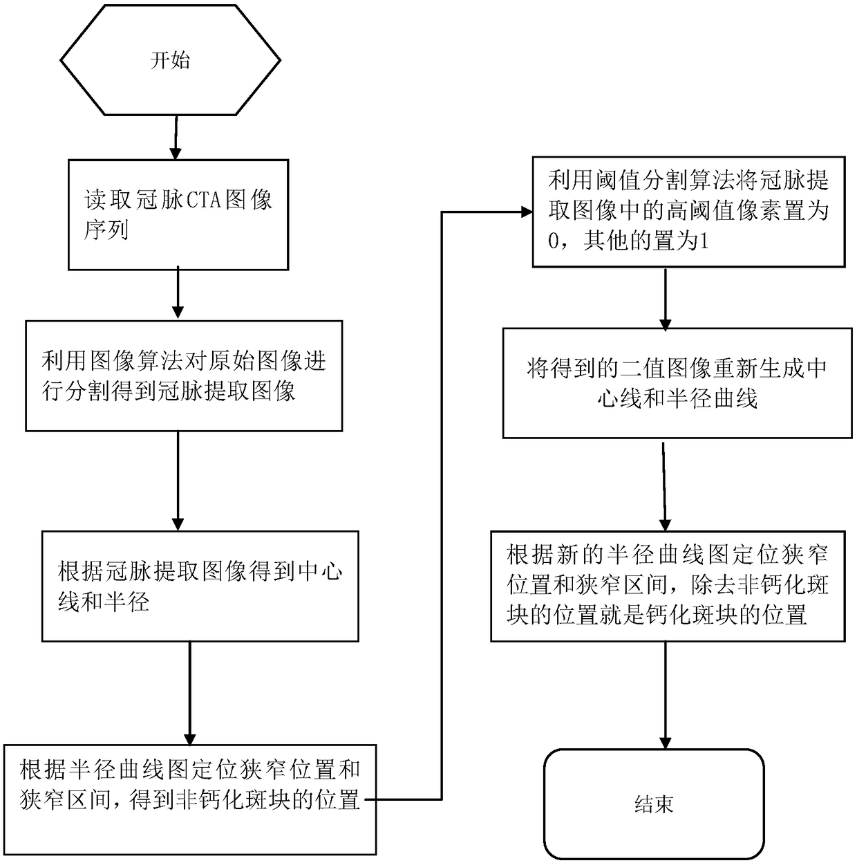
[0024] Such as figure 1 As shown, the plaque classification method for coronary CT images of the present invention includes the following steps.

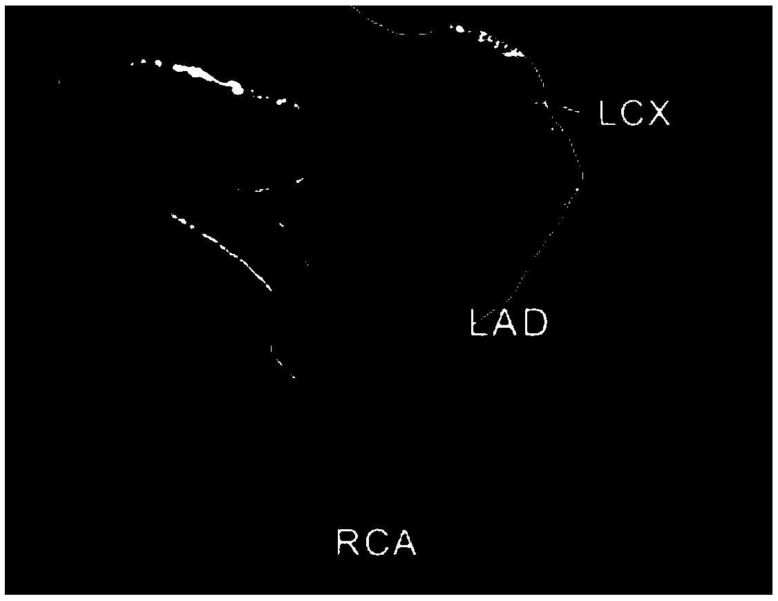
[0025] Step S1: Carry out coronary artery segmentation on the CT image to obtain a coronary artery extraction map (with calcified plaque and stenosis).

[0026] Read the CT image data, first frame the heart area to exclude bone interference, and obtain the heart extraction im...


PUM


Abstract

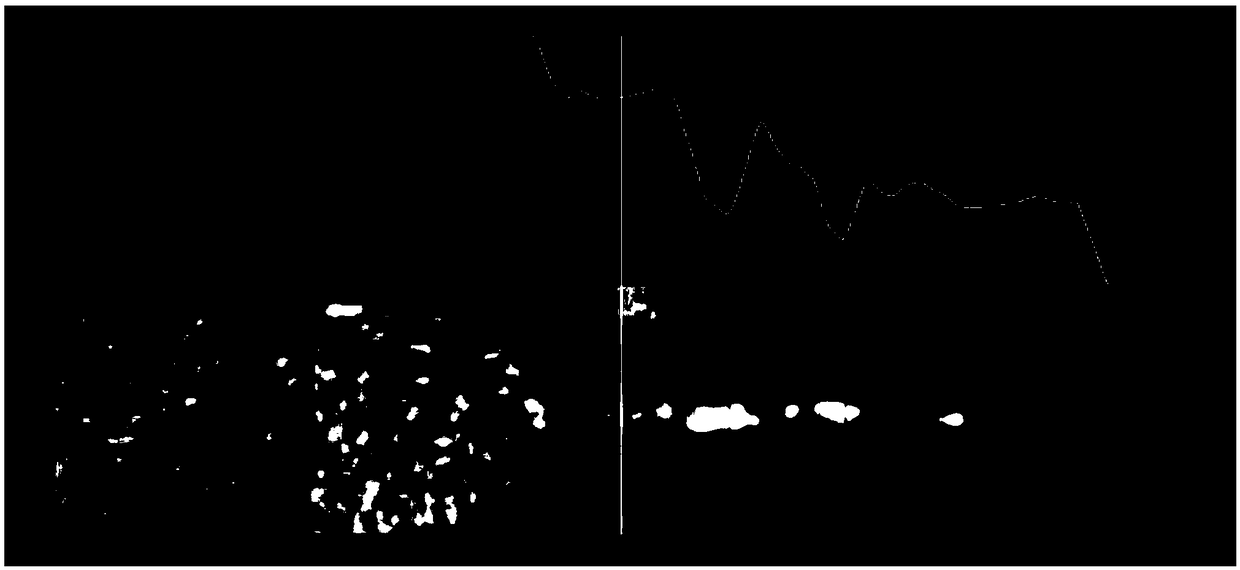
The invention discloses a plaque classification method for coronary CT images. The method comprises: segmenting a coronary CTA sequence original image to obtain a coronary extraction map; extracting an coronary tree data isosurface, generating grid model data, calculating a normal vector, locating the start and end points from all points on a grid model, calculating the shortest path between the start point and the end point, and equidistantly filtering an obtained curve to obtain a center line and a radius; locating a bright spot in the coronary extraction map; locating a stenosis position and a stenosis section according to the radius and the curve; obtaining a coronary binary image with calcified plaque removed, obtaining the center line of the coronary binary image with calcified plaque removed, and calculating the radius; locating the stenosis position andthe stenosis section according to the radius and the curve, and determining that the plaque is a non-calcified plaque if the stenosis position is the same as the stenosis position in the step S04, otherwise determining that the plaque is a calcified plaque. The method can detect calcified and non-calcified plaques in CTA images and automatically classifies the plaques.

Description

technical field [0001] The invention relates to the technical field of medical image processing, in particular to a plaque classification method for coronary CT images, which can be applied to X-ray coronary angiography image analysis in clinical research. Background technique [0002] Finding a safe and reliable method for detecting coronary artery disease is the main goal of clinical development in the future, so it is of great clinical value and practical significance to be able to accurately extract plaque from CTA image sequences to judge coronary artery disease. In the past ten years, the proportion of deaths caused by coronary artery disease has been increasing year by year. Therefore, it is necessary to accurately extract the quantification of arterial vascular disease, especially the early detection and quantitative analysis of plaque. However, the early detection and quantification of plaque requires very experienced doctors to spend a long time on manual plaque se...

Claims


Application Information

Patent Timeline
IPC IPC(8): G06T7/00G06T7/62G06T7/68G06T7/11G06T7/136
CPCG06T7/0012G06T2207/10081G06T2207/30101G06T7/11G06T7/136G06T7/62G06T7/68
Inventor 霍云飞王之元曹文斌张海玲
Owner SUZHOU RAINMED MEDICAL TECH CO LTD
Learn More