
Digital medical X-ray photography system

An X-ray and X-ray tube technology, applied in the field of digital medical X-ray photography system, can solve the problems of narrow application range of ordinary seats, discount of patient comfort, secondary injury of feet, etc., to reduce the risk of secondary injury, Check the effect of ease of operation and pain reduction

Active Publication Date: 2018-12-18
孙媛媛
View PDF13 Cites 6 Cited by
  • Summary
  • Abstract
  • Description
  • Claims
  • Application Information

AI Technical Summary

Problems solved by technology

The detector of the existing digital medical X-ray photography system still has a certain height from the ground at the lowest position of the stand. Because the patient's ankle has trauma, when the position is set, the patient usually sits on the seat and stands on the ground. With the assistance of the physician, the injured foot is placed on the detector. Due to the different heights and shapes of patients, such as children and adults, the scope of application of ordinary seats is relatively narrow, and the comfort experienced by patients is also greatly reduced.
In order to better observe the gaps between the small joints of the foot, the patient needs to apply force to the foot under the guidance of the doctor. It is inconvenient to apply force in the previous sitting posture, and improper force application in the standing position will cause secondary damage to the patient's foot. secondary injury, aggravating the pain of the patient

Method used


Image

  • Digital medical X-ray photography system
  • Digital medical X-ray photography system
  • Digital medical X-ray photography system

Examples


Embodiment 1

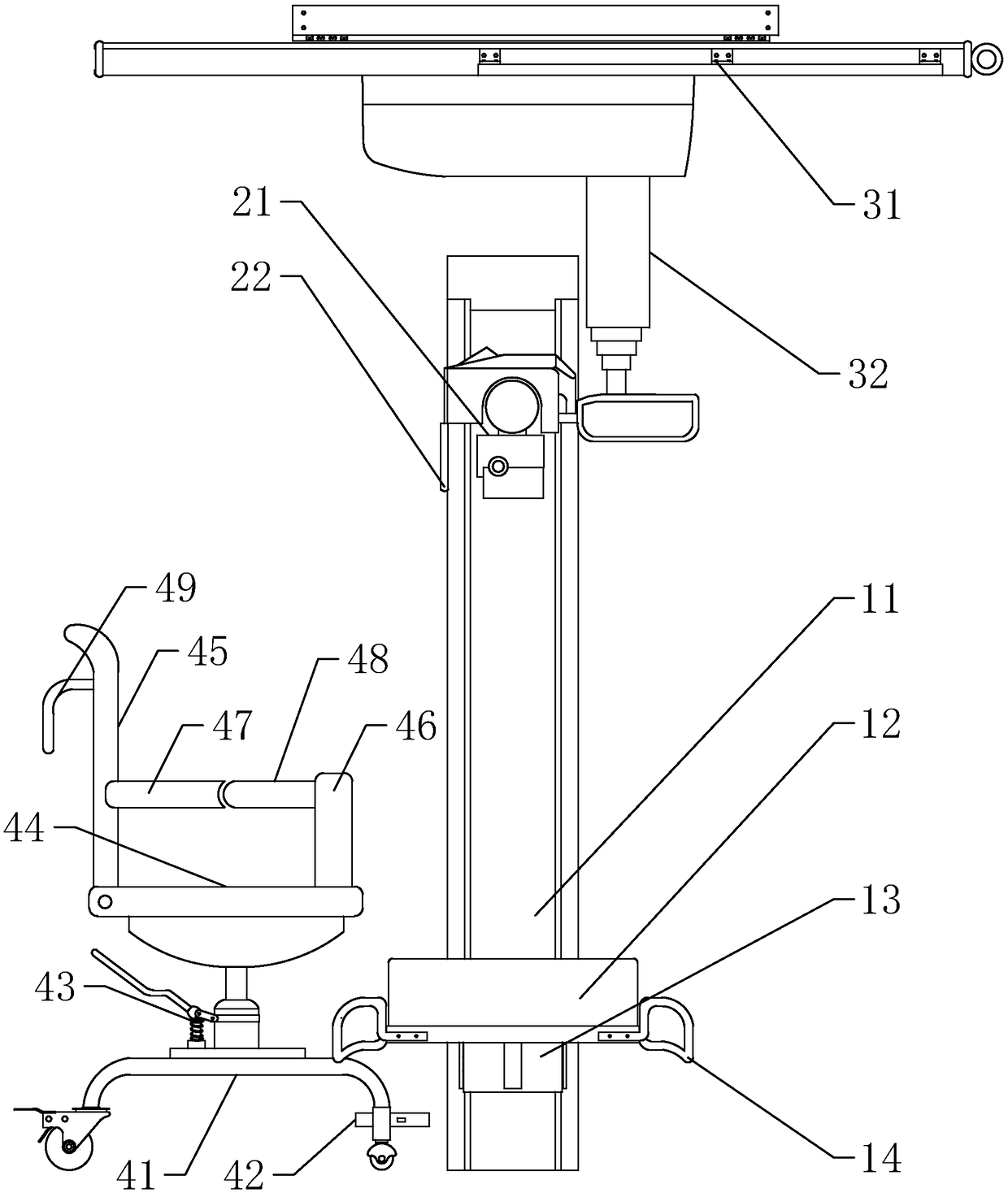
[0025] Such as figure 1 As shown in , the digital medical X-ray imaging system of this embodiment includes a vertical photographic frame, a detector, an X-ray tube assembly, and an X-ray tube assembly bracket, wherein the vertical photographic frame includes a column 11, which is rotatably connected with a detector 12 The detector tray 13 can move up and down along the column 11. There are control panels and handles 14 on both sides of the detector 12. Through the control panel, the detector 12 can be controlled to tilt to a horizontal position or to a vertical position and to detect The detector tray 13 moves up and down, and the movement of the detector 12 can also be manually adjusted through the handle 14; the detector 12 converts the X-ray energy into a digital signal of the image, and the strength of the ray signal received by the detector depends on the position The density of tissues in the human body section finally forms images with different grayscales; the X-ray tu...

Embodiment 2

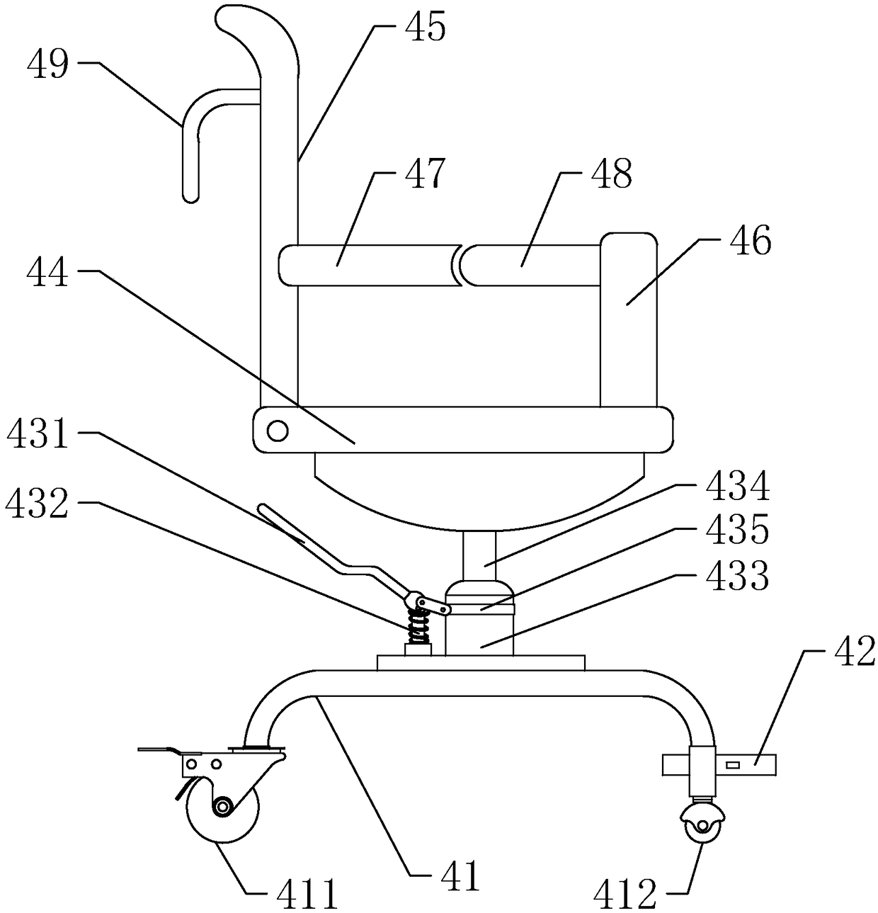
[0032] Such as Figure 8 and 9 As shown, the difference between this embodiment and the first embodiment above is that in this embodiment, no friction ring is provided at the connection between the bottom frame 41 and the rotating shaft 421, but a linkage locking mechanism is provided on the pedal 42 , the linkage locking mechanism includes a U-shaped plug-in 422, the two free ends of the U-shaped plug-in 422 cooperate with the corresponding locking sockets on the chassis 41, and the rear end of the U-shaped plug-in 422 is connected to the pedal 42 through a linkage rod 423. The linkage button 424 on the side is connected, and a return spring 425 is provided between the linkage button 424 and the pedal 42 .

[0033] During use, by controlling the insertion of the U-shaped plug-in 422 and the locking jack, the horizontal position of the pedal 42 is locked. When the seat is pushed to the detector 12, the linkage button 424 is pressed inward, and the linkage lever 423 will drive...


PUM


Abstract

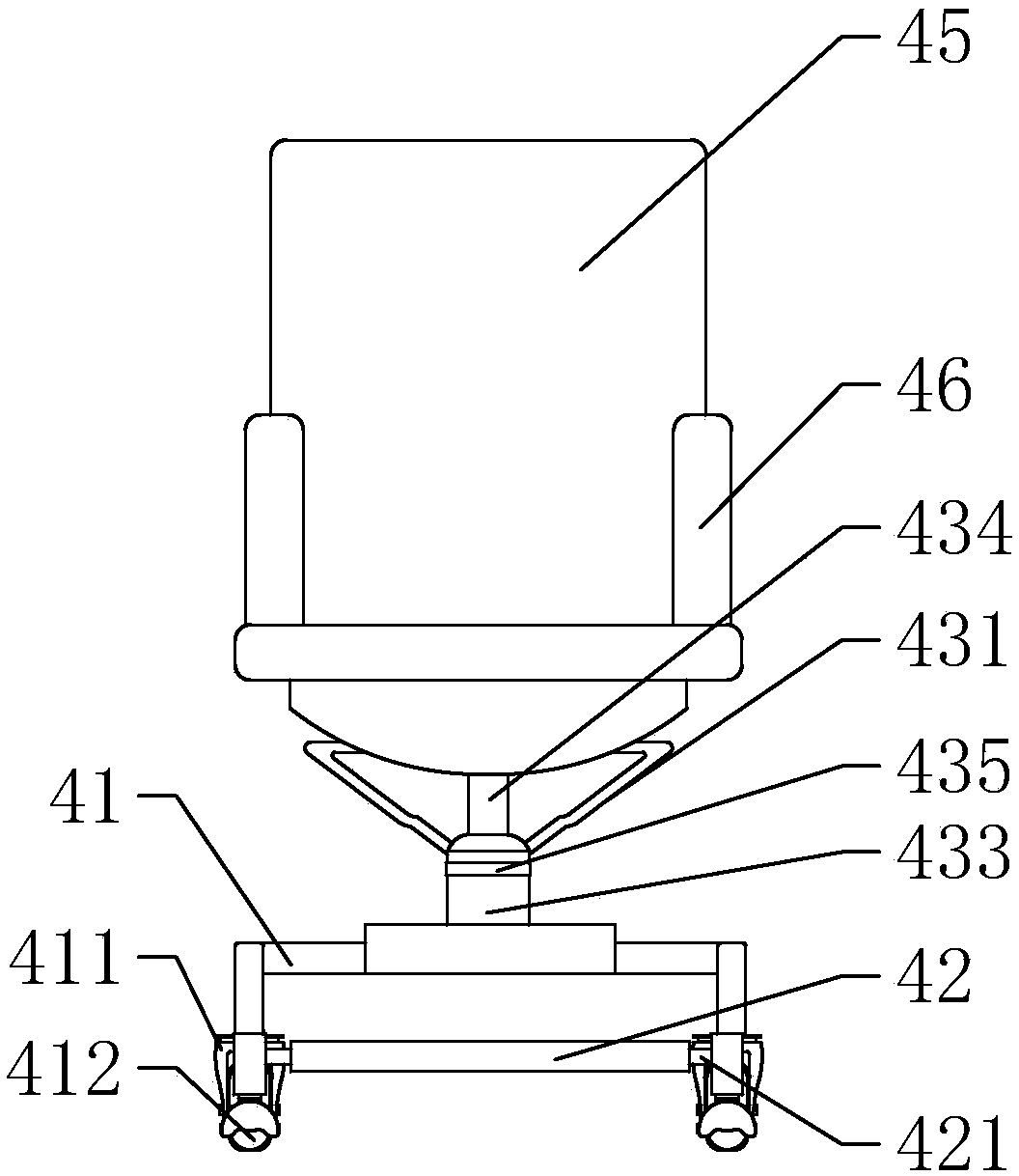
The invention discloses a digital medical X-ray photography system. The system includes an upright frame, a detector, an X-ray tube module, an X-ray tube module support and a seat with a pressure applying device. The seat is provided with trundles, a foot rest and a hydraulic step-lifting device. A piston of the hydraulic step-lifting device is connected with a bearing seat plate. The bearing seatplate is provided with a seat back and a support frame. Two sides of the seat back are respectively provided with fixed handrails, and the pressure applying device is arranged in the support frame. Amovable armrest is connected with one end of a pressing upright rod through a rotating pair, and the other end of the pressing upright rod forms a moving pair with a slide way fixed on the bearing seat plate. The pressing device also comprises a ratchet mechanism which is mainly composed of a pawl and a ratchet rack fixed on the support frame. The X-ray photography system of the invention is provided with the dedicated supporting seat, which is convenient for checking operation and enhances the comfort level. Safety and reliability are improved and the pain suffered by a patient when the patient needs positioning is reduced, and risks of secondary injury are reduced.

Description

Technical field: [0001] The invention relates to a medical device, in particular to a digital medical X-ray photography system. Background technique: [0002] X-ray diagnosis is one of the important clinical diagnostic methods, which is based on X-ray images. X-ray images are composed of images with different grayscales from black to white, and are grayscale imaging. These images of different gray levels reflect the anatomy and pathological state of human tissue structure with density. In recent years, with the improvement of medical equipment level, digital medical X-ray photography system has been widely used to improve the level of diagnosis and treatment. Digital medical X-ray photography system is a radiographic equipment using direct digital conversion technology software, which includes direct imaging digital flat panel detectors, radiation generating devices and automatic image acquisition and post-processing software. They form an image chain, which can provide cl...

Claims


Application Information

Patent Timeline
Patent Type & Authority Applications(China)
IPC IPC(8): A61B6/00
CPCA61B6/00
Inventor 孙媛媛刘德华徐莉丽刘朔闻赵若妤
Owner 孙媛媛
Learn More