A nerve fiber model for quality control of magnetic resonance diffusion imaging and its preparation method
A technology of imaging quality and nerve fiber, applied in application, diagnostic recording/measurement, medical science, etc., can solve the problems of long cycle, complicated model making process, no physical model of diffusion imaging fiber, etc., and achieve good simulation effect, The water filling process is convenient and quick
- Summary
- Abstract
- Description
- Claims
- Application Information
AI Technical Summary
Problems solved by technology
Method used
Image

Examples
Embodiment 1
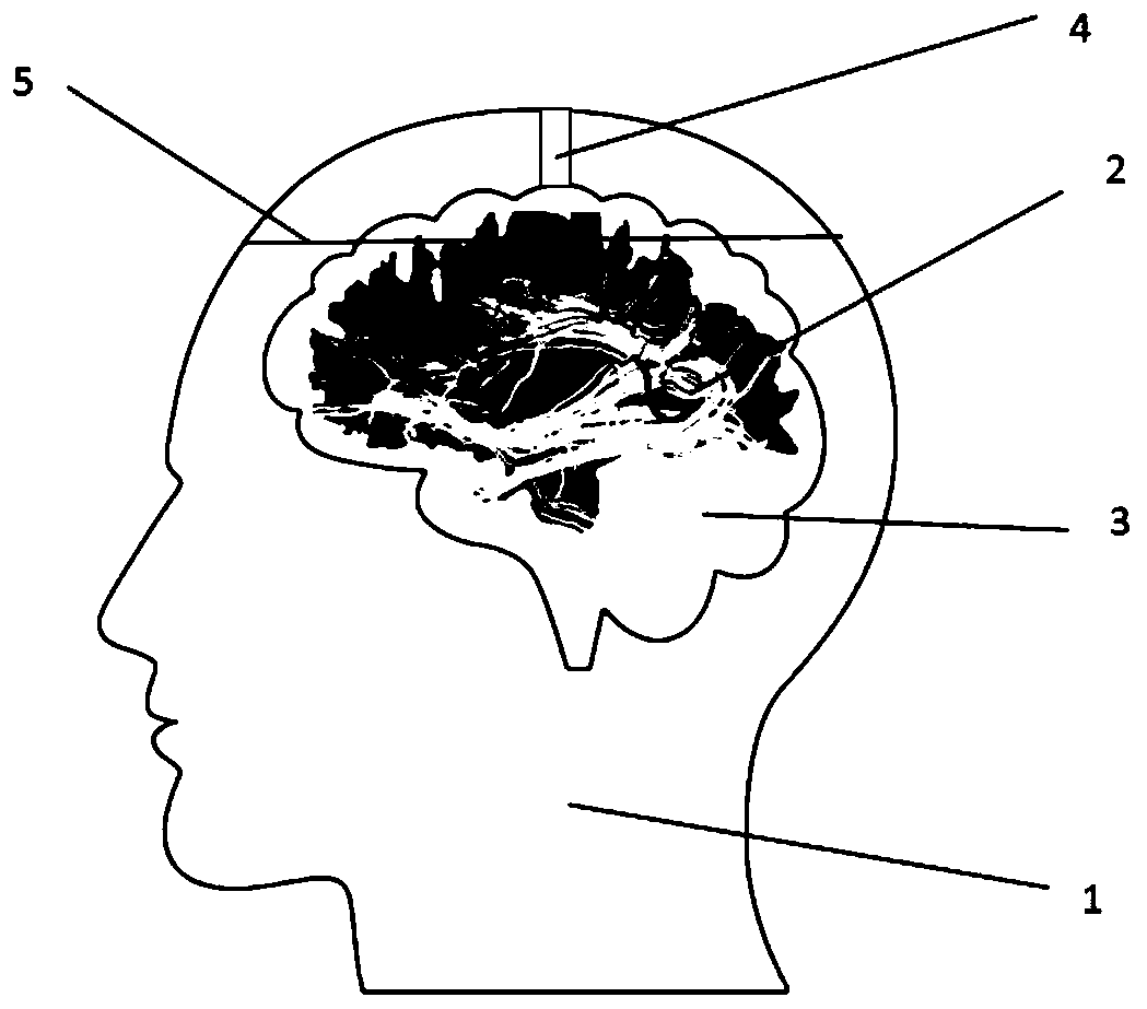
[0031] Firstly, prepare the 3D printed braincase: use the 3T magnetic resonance imaging system T1-mprage sequence to scan the head of healthy human subjects, and obtain the 3D T1 structure image with f resolution of 1mm*1mm*1mm. Then Minics software was used for graphic segmentation, brain tissue was removed from the original image, and the hollow braincase structure was obtained by smoothing. Then convert the brain structure map into a .stl file that can be recognized by 3D printing. Then divide the 3D head-shell diagram into two parts that can be covered up and down along the dividing line 5. The upper part has a shorter distance from the top of the head and the lower part has a larger opening. A glue injection hole 4 with a diameter of 5-10mm. Print the modified .stl file with PLA or SLA material to obtain the skull structure 1, such as figure 1 shown.
[0032]Preparation of spinning: the wall material of the electrospinning fiber is a mixture of PCL and PVP in a mass ra...
Embodiment 2
[0035] The preparation method of the 3D printed braincase is the same as in Example 1, wherein the 3D T1 structure image is obtained by scanning with a 7T magnetic resonance imaging system with a resolution of 0.5mm*0.5mm*0.5mm.
[0036] The preparation of spinning is the same as in Example 1.
[0037] The preparation of the agarose gel and the fixation of the spinning were the same as in Example 1, in which a part of the agar had a mass concentration of 2% for simulating fibrotic brain tumors, and the other agar had a mass concentration of 1.5% for simulating conventional brain tissue. The preparation method is to firstly prepare 2% agar, remove excess gel after the agar is solidified, and retain the desired shape and size of the gel block of the brain tumor. On this basis, electrospun fibers and 1.5% agar were further added.
[0038] All the other steps are the same as in Example 1.
Embodiment 3
[0040] In this example, 0.1 mmol / L manganese chloride and 0.2 mmol / L nickel chloride were added to the agarose gel in Example 1, so that the T1 and T2 values of the agar were closer to human gray matter. In addition, sodium nitrite, an antifungal substance with a mass concentration of 30 mg / kg, was added.
PUM
 Abstract
Description
Claims
Application Information
 - R&D Engineer
- R&D Manager
- IP Professional
- Industry Leading Data Capabilities
- Powerful AI technology
- Patent DNA Extraction
Browse by: Latest US Patents, China's latest patents, Technical Efficacy Thesaurus, Application Domain, Technology Topic.
© 2024 PatSnap. All rights reserved.Legal|Privacy policy|Modern Slavery Act Transparency Statement|Sitemap