
A convolution neural network method for differentiation of hepatocellular carcinoma

A convolutional neural network and liver cancer technology, applied in the super-field, can solve the problems of missed diagnosis, time-consuming, etc., and achieve the effect of accurately distinguishing results, avoiding strong observation, and avoiding shortcomings and missed diagnosis.

Inactive Publication Date: 2019-01-15
FUJIAN NORMAL UNIV
View PDF5 Cites 4 Cited by
  • Summary
  • Abstract
  • Description
  • Claims
  • Application Information

AI Technical Summary

Problems solved by technology

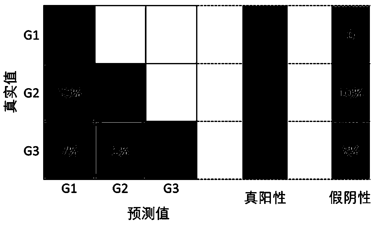
[0005] The purpose of the present invention is to provide a convolutional neural network method for distinguishing liver cancer differentiation grades, which can effectively overcome the shortcomings of time-consuming, missed diagnosis, and strong subjectivity in current clinical biopsy techniques

Method used


Image

  • A convolution neural network method for differentiation of hepatocellular carcinoma
  • A convolution neural network method for differentiation of hepatocellular carcinoma
  • A convolution neural network method for differentiation of hepatocellular carcinoma

Examples


Embodiment Construction

[0015] The technical solution of the present invention will be specifically described below in conjunction with the accompanying drawings.

[0016] The present invention provides a method for distinguishing the differentiation grade of liver cancer by a convolutional neural network, which is used to distinguish the MPM image data of liver cancer biopsy samples or samples sent for inspection after liver cancer resection, including the following process:

[0017] 1. Prepare data;

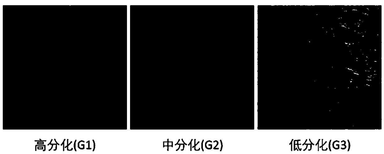
[0018] 217 MPM images of liver cancer were collected by a multiphoton microscope system (eg figure 2 ), set the excitation wavelength of the system to 810nm, and select an upright 20× objective lens (0.8 NA) to scan the sample. Set up 2 receiving channels, one at 370-419 nm, marked in green, for collecting SHG signals; the other at 420-700 nm, marked in red, for detecting TPEF signals. The size of a single image is 425.10×425.10 μm2, and the pixels are 512×512. Diagnosed by experienced pathologist...


PUM


Abstract

The invention relates to a method for distinguishing differentiation grade of liver cancer by convolution neural network. The method comprises the following steps: step S1, preparing data: acquiring MPM images of liver cancer through a multi-photon microscope; 2, enlarging the MPM image data set of the liver cancer: adjusting the pixel of the original pixel size of 512x512 collected in the step S1, rotating the image horizontally, rotating vertically symmetrically, cutting and the like, so as to obtain the image number of the training set to be 16 times of the original image number; S3, designing a convolution neural network to obtain the differentiation result of the differentiation grade: designing the structure of the convolution neural network. The convolution neural network consists of eight layers: the first layer to the fifth layer is called convolution layer, which is used to extract the detailed features from the image; layer 6 to layer 8 are three fully connected layers, andafter layer 8, the probability of differentiation grade of liver cancer image is output. The invention can effectively overcome the shortcomings of time-consuming, missed diagnosis and strong subjectivity in the current clinical biopsy technology.

Description

technical field [0001] The invention belongs to the field of medical image processing, and in particular relates to a convolutional neural network method for distinguishing liver cancer differentiation grades. Background technique [0002] Liver cancer is the most common fatal malignancy, with 466,100 new cases and 422,100 deaths in 2015 in China alone. Poorly differentiated HCC has strong aggressiveness and poor prognosis, resulting in a low 5-year survival rate. Liver cancer patients with different grades of tumor differentiation correspond to different prognosis and treatment strategies, and have completely different results. Therefore, the differentiation of HCC differentiation grades has great clinical value. At present, commonly used methods, such as histopathological examination and B-ultrasound, are easily affected by the experience and discrimination of examiners. It is necessary to develop a new method to achieve label-free, rapid, quantitative and automatic dif...

Claims


Application Information

Patent Timeline
IPC IPC(8): G06K9/62G06N3/04
CPCG06V2201/032G06N3/045G06F18/241G06F18/214
Inventor 林宏心韦超王光兴卓双木
Owner FUJIAN NORMAL UNIV
Learn More