A microscopic image analysis method of a cervical cancer tissue based on a graph theory
A technology of microscopic images and analysis methods, applied in the field of image analysis, can solve problems such as affecting the structure, and achieve the effect of enhancing the accuracy, speeding up the time of diagnosis, and improving the accuracy of diagnosis
- Summary
- Abstract
- Description
- Claims
- Application Information
AI Technical Summary
Problems solved by technology
Method used
Image
Examples
Embodiment Construction
[0043] In order to better explain the present invention and facilitate understanding, the present invention will be described in detail below through specific embodiments in conjunction with the accompanying drawings.
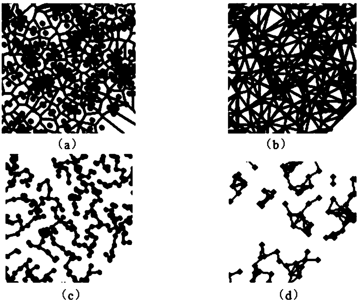
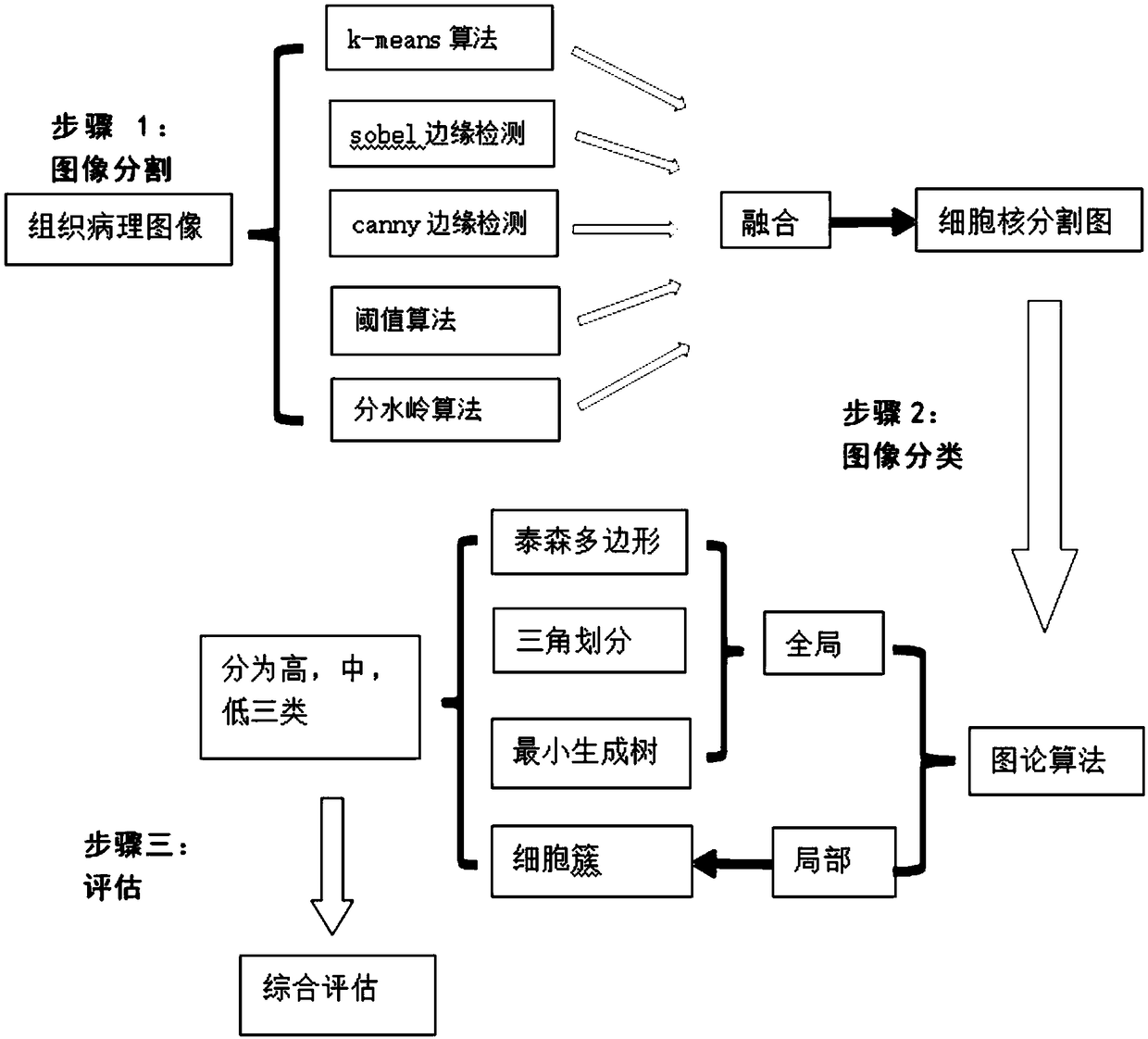
[0044] refer to image 3 , in this embodiment, a method for analyzing microscopic images of cervical cancer tissue based on graph theory is provided, which includes the following steps:
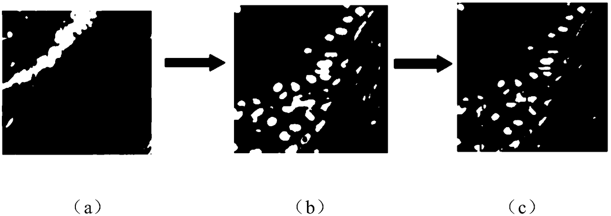
[0045] Step A: collect microscopic image data of cervical cancer tissue, use different algorithms to segment each original image collected, and fuse the segmentation results to obtain a fused image.
[0046] In step A, the original image formats collected include *.bmp, *.BMP, *.dip, *DIP, *.jpg, *.JPG, *.jpeg, *JPEG, *.jpe, *.JPE, *.jfif, *JFIF, *.gif, *.GIF, *.tif, *.TIF, *tiff, *.tiff, *.png, *.PNG, etc.: For example, the experimental dataset used in this patent contains 360 images, each image size is 2560x1920 pixels.
[0047] Segmenting each original image collected in...
PUM
 Abstract
Description
Claims
Application Information
 - R&D
- Intellectual Property
- Life Sciences
- Materials
- Tech Scout
- Unparalleled Data Quality
- Higher Quality Content
- 60% Fewer Hallucinations
Browse by: Latest US Patents, China's latest patents, Technical Efficacy Thesaurus, Application Domain, Technology Topic, Popular Technical Reports.
© 2025 PatSnap. All rights reserved.Legal|Privacy policy|Modern Slavery Act Transparency Statement|Sitemap|About US| Contact US: help@patsnap.com


