Multi-stage collagen-based template or implant for use in the repair of cartilage lesions

Inactive Publication Date: 2000-06-27
HOSPITAL FOR JOINT DISEASES ORTHOPEDIC INST
View PDF8 Cites 309 Cited by
  • Summary
  • Abstract
  • Description
  • Claims
  • Application Information

AI Technical Summary

Benefits of technology

It is a further object of this invention to provide a multi-staged collagen implant to repair cartilage lesions which is effective and safe.
It is another object of this invention to provide a multi-staged collagen implant to repair cartilage lesions which is resorbable.
Defects in articular cartilage can be healed by utilizing a regeneration template formed by combining a porous collagen sponge ("collagen matrix") with a dense collagen membrane. The dense collagen membrane is made with a pore size of less than 1 um (micrometer), and is cross-linked with a non-cytotoxic agent to increase strength and lengthen resorption time. Be

Problems solved by technology

It has been well documented that injured articular cartilage has only a limited ability for self-repair.
As articular cartilage is relatively avascular and aneural, loss of the surface tissue will result in a permanently scarred site.
Current treatment protocols call for surgical intervention (such as abrasion arthroplasty, excision and drilling, articular cartilage debridement, and arthroscopic shaving) and will most often lead again to inadequate repair.
Long-term morbidity such as degeneration to arthritic conditions will often result in patients with chronic cartilage problems.
These techniques have been used to transplant whole or partial joints, with mixed results.
Although these approaches gave repair tissue that more closely resembled normal car

Method used


Image

  • Multi-stage collagen-based template or implant for use in the repair of cartilage lesions
  • Multi-stage collagen-based template or implant for use in the repair of cartilage lesions
  • Multi-stage collagen-based template or implant for use in the repair of cartilage lesions

Examples


Example

EXAMPLE 2

Preparation and Characteristics of the Dense Collagen Membrane

A dense collagen membrane is prepared according to the procedure presented in U.S. Pat. No. 5,206,028, the entire disclosure of which is incorporated by reference herein. A porous matrix, having a thickness of 4 mm to 10 mm, is hydrated using a humidity controlled chamber, relative humidity of 80% at 25.degree. for 60 minutes. The moist collagen material is compressed between two Teflon sheets to a thickness of less than 0.2 mm. The compressed material is then cross-linked in a solution of 0.5% formaldehyde, 1% sodium bicarbonate at pH 8 to 60 minutes. The cross-linked membrane is then raised thoroughly with water, then freeze dried overnight under similar conditions as in Example 1, except that the time for freeze drying is about 48 hours. The dense collagen membrane has an inner construction of densely packed fibers that are intertwined in a mufti-layer structure. The collagen membrane allows diffusion of molec...

Example

EXAMPLE 3

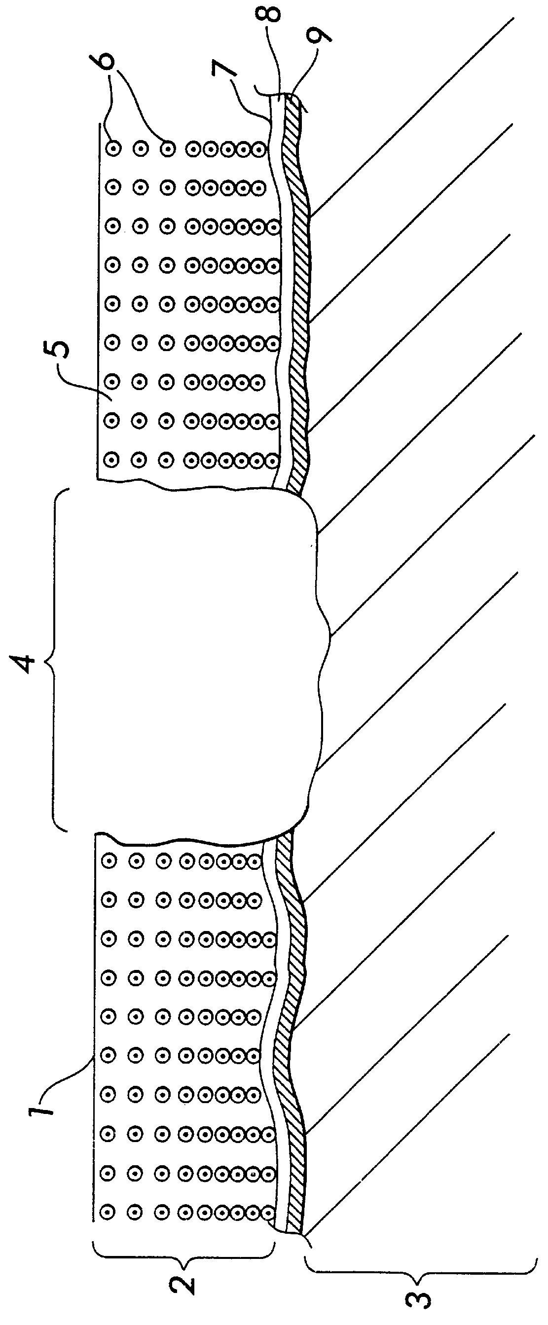
Utilization of Non-cell Seeded Matrix in Cartilage Defects

1. Prepare a surgically defined site, slightly smaller than the size of the matrix implant. The depth should be approximately the same size of the collagen matrix / cell composite. The surgically prepared site should go through the subchondral plate (i.e., a bleeding bed).

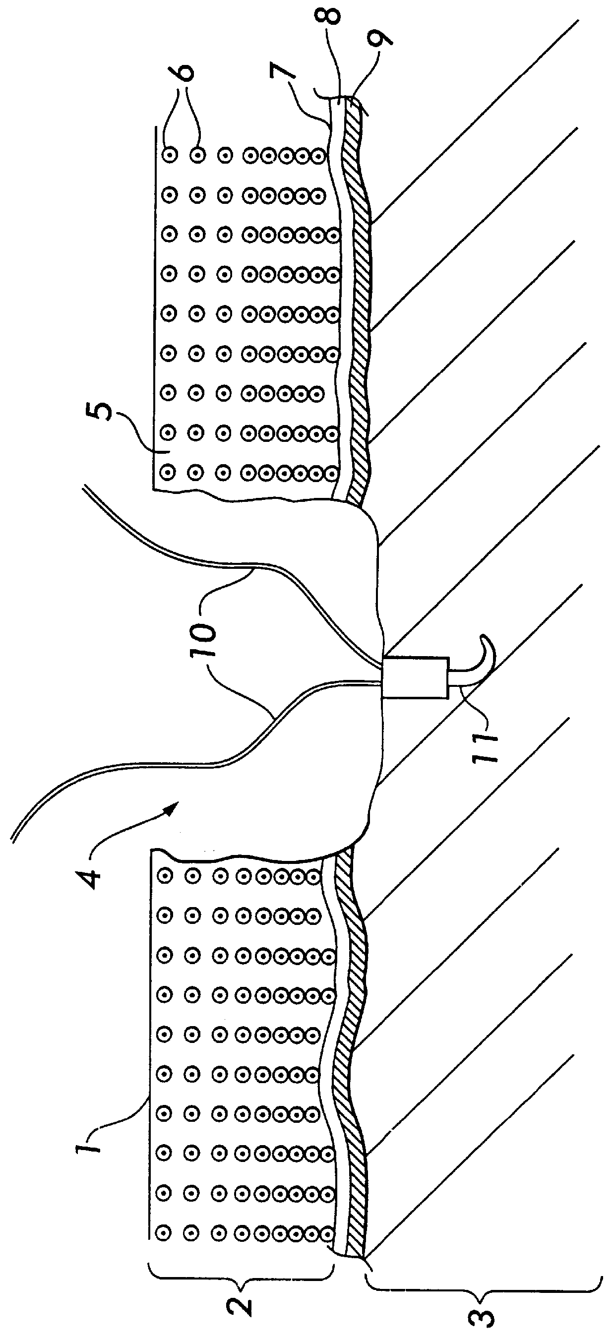
2. Set an anchor, with two attached resorbable suture lines, into the center of the surgically prepared site (FIG. 2). There will be four lines available, one in each quadrant.

3. Attach a dense collagen membrane (pore structure less than one micrometer) onto the collagen matrix, using resorbable sutures such as Vicryl 7-0 (FIG. 3).

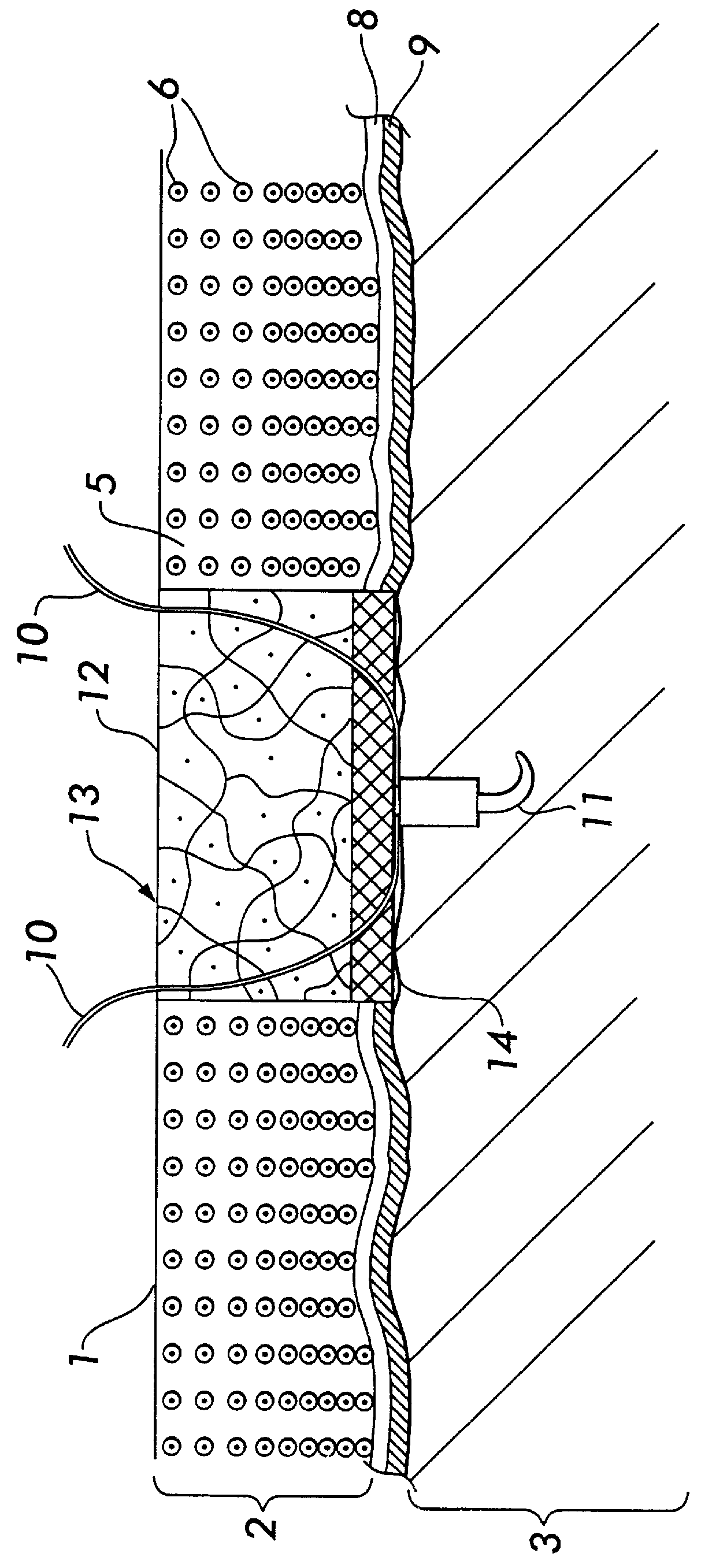
4. Place the template with the dense collagen membrane on the bottom of the surgically prepared site, securing it in place with the suture lines by threading the suture lines through the template.

5. Place a protecting piece of tibial periosteum over the matrix. The periosteum must be oriented over the implant as follows. The...

Example

EXAMPLE 4

Utilization of Cell-Seeded Matrix in Cartilage Defects

1. Obtain autologous sample of tissue containing cartilage or progenitor cells.

2. Remove extracellular matrix from the tissue sample, then isolate cells using standard methods.

3. Expand cells in culture.

4. The dense collagen membrane is attached to the collagen matrix with bioresorbable sutures, such as Vicryl 7-0.

5. Add cells to the pore-defined collagen matrix, so that cells penetrate through the matrix. This can be done by laying the cell suspension over the matrix, then carefully applying a vacuum under the matrix.

6. Culture the collagen template / cell composite for one week or more.

7. Prepare a surgically defined site, slightly smaller than the size of the implant. The depth should be approximately the same size of the collagen template / cell composite. The surgically prepared site should go through the subchondral plate (i.e., a bleeding bed).

8. Set an anchor, with two attached resorbable suture lines, into the cente...


PUM

PropertyMeasurementUnit
Pore sizeaaaaaaaaaa
Pore sizeaaaaaaaaaa
Thicknessaaaaaaaaaa

Abstract

The invention is a template to aid in the regeneration of articular cartilage. The template is formed by combining a porous collagen sponge ("collagen matrix") with a dense collagen membrane. The dense collagen membrane is placed on the surface of the cartilage defect to prevent cell migration from the subchondral plate and vasculature. The collagen membrane will allow movement and exchange of fluids, nutrients, cytokines and other factors necessary for cartilage regeneration. The collagen matrix has been developed to allow attachment and growth of cells, specifically chondrocytes which are normally found in articular cartilage. The collagen matrix can be combined with chondrocytes in vitro, and therefore serve to transport cultured cells to the defect site and to retain the cells in position following implantation. Procedures are described to effectively use the two-staged template, and to fix the template to the repair site.

Description

This invention is within the technical fields of surgery, medicine, tissue engineering, biology, biomaterials, polymers, and biochemistry. It is both a product and a method of use for the repair of cartilage lesions, with and without the use of chondrocytes cultured in vivo.It has been well documented that injured articular cartilage has only a limited ability for self-repair. As articular cartilage is relatively avascular and aneural, loss of the surface tissue will result in a permanently scarred site. Lesions which fracture the subchondral bone which has a greater vascular supply will undergo an inflammation / repair response, with the damaged site filling with fibrocartilage tissue (Convery, et al. 1972). In either case, function is impaired and chronic pain is the usual prognosis as the biochemical and biomechanical characteristics of the cartilage have been altered. Current treatment protocols call for surgical intervention (such as abrasion arthroplasty, excision and drilling, ...

Claims


Application Information

Patent Timeline
IPC IPC(8): A61F2/28A61F2/30A61L27/00A61L27/24A61F2/02A61F2/00A61B17/04A61B17/00A61B17/06
CPCA61F2/2846A61F2/30756A61L27/24A61B17/0401A61L2430/06A61B2017/00004A61F2002/2835A61F2002/30013A61F2002/30062A61F2002/30461A61F2002/30682A61F2002/30751A61F2002/30761A61F2210/0004A61F2220/0075A61F2250/0024A61F2310/00365A61B17/06166A61F2002/30011
Inventor PACHENCE, JAMES M.FRENKEL, SALLYMENCHE, DAVID
Owner HOSPITAL FOR JOINT DISEASES ORTHOPEDIC INST
Why Eureka
Try Eureka