Implantable medical device for treating cardiac mechanical dysfunction by electrical stimulation

a technology of electrical stimulation and medical devices, applied in the field of implantable medical devices, can solve the problems of slow response time, and achieve the effect of minimizing the potential risk of shock-induced myocardial damage and high detection specificity

Inactive Publication Date: 2006-08-22
MEDTRONIC INC
View PDF32 Cites 327 Cited by
  • Summary
  • Abstract
  • Description
  • Claims
  • Application Information

AI Technical Summary

Benefits of technology

[0042]Combined with negative inotrope drug treatments such as beta blockers to improve patient tolerance of these treatments;
[0098]A second change is to increase the time between the later shocks in the sequence. With greater spacing, higher detection specificity would be possible and minimize the potential risk of shock-induced myocardial damage.

Problems solved by technology

This may be thought of as closing the loop but results in a slow response time.
In this patent disclosure, the inventors report that they increased the controller gain to the point where oscillations developed, an instability phenomenon well known in the area of feedback control.

Method used


Image

  • Implantable medical device for treating cardiac mechanical dysfunction by electrical stimulation
  • Implantable medical device for treating cardiac mechanical dysfunction by electrical stimulation
  • Implantable medical device for treating cardiac mechanical dysfunction by electrical stimulation

Examples


example 1

AED Example with Presentation of VF

[0238]Despite the increasing availability to quick access defibrillation by the public and quickening response times, the prognosis of a victim of a sudden cardiac arrest surviving to a hospital discharge is low, with many of these victims succumbing to electromechanical dissociation (EMD) or pulseless electrical activity (PEA). Current AED technology is equipped to treat tachyarrhythmias but has no means to treat EMD / PEA.

[0239]An AED equipped with the features detailed in this invention would address these scenarios. In an example implementation, the AED would appear identical to the first responder. The responder would place two transthoracic self-adhesive electrodes on the patient and depress a start button on the device. The AED would then obtain a surface ECG from the transthoracic electrodes and apply a VF detection algorithm to the signal. If VF was detected, the AED would apply a defibrillation shock and then apply a re-detection algorithm....

example 2

ICD Example with Presentation of VT

[0241]ICD systems provides patients with greatly improved survivability from episodes of sudden cardiac arrest when compared to patients treated with AED's mainly because there is minimal time to wait between the onset of the arrhythmia and delivery of therapy when the device is implanted and always ready to detect events. However, some patients, especially those with more pronounced HF, may not tolerate well even the shortest of VF episodes and may have depressed cardiac function long after the arrhythmia is terminated. Additionally, circumstances may arise that lengthen the duration of the tachyarrhythmia before the device delivers a therapy. Some tachyarrhythmias pose detection problems for ICD's, which may postpone delivery of therapy. An arrhythmia could also require several shocks to terminate, further prolonging the episode.

[0242]During a tachyarrhythmia, the coronary blood flow perfusing the heart can become severely impaired, leading to is...

example 3

HF Example with Presentation of Acute Decompensation

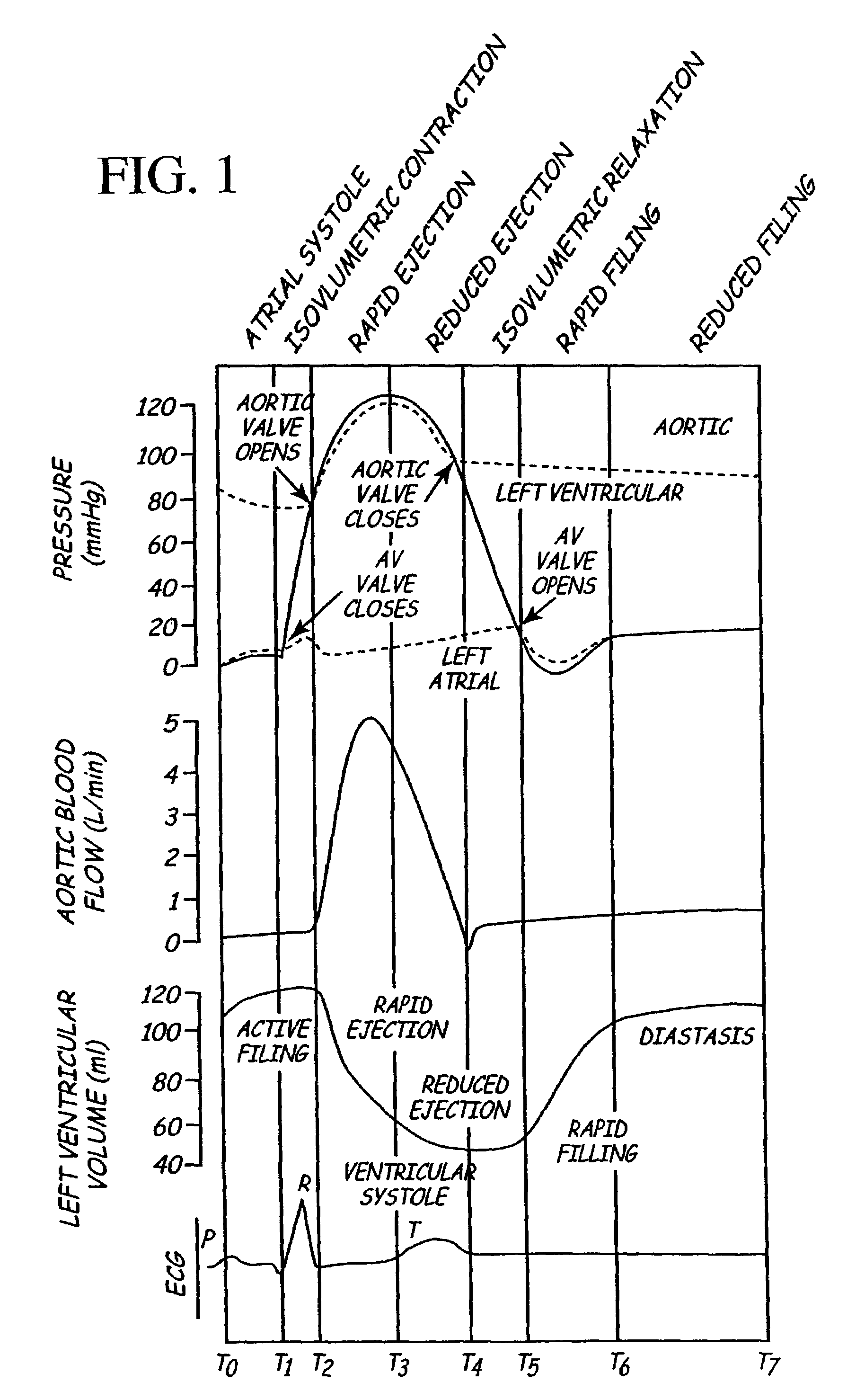
[0244]Advanced stage HF patients experience sudden worsening of heart failure associated symptoms which require hospitalization. The transition from chronic compensated HF to acute decompensated HF may result from a number of factors including dietary indiscretion, progress of HF disease, and acute myocardial infarction. When severe, symptoms may progress in a few hours to a stage where these patients need to be admitted to a critical care hospital bed, monitored by physiologic sensors, and treated with a variety of drugs including intravenous inotropes. A patient experiencing such a decompensation commonly exhibits low cardiac output at rest, poor contractile function and low dP / dt max, slow relaxation and high tau, elevated diastolic ventricular pressures, and reduced ventricular developed pressures.

[0245]Cardiac resynchronization therapy delivered by an implanted device is an important adjunct to good medical therapy. Such a res...


PUM


Abstract

The disclosure provides methods and apparatus of particular benefit for patients suffering heart failure including cardiac dysfunction, chronic HF, and the like and all variants thereof. According to the disclosure monitoring and therapy delivery for a wide variety of acute and chronic cardiac dysfunctions are described and depicted. Various forms of paired or coupled pacing therapy delivery provided alone or in combination with neurostimulation therapy delivered by both implantable and external apparatus, including defibrillation therapy are also provided herein.

Description

CROSS-REFERENCE TO RELATED APPLICATIONS[0001]This patent disclosure claims the benefit of provisional U.S. Patent Application Ser. No. 60 / 315,316 filed 28 Aug. 2001 the entire contents of which are hereby incorporated by reference herein.[0002]This patent disclosure hereby incorporates by reference commonly assigned U.S. Pat. No. 6,438,408 which issued 20 Aug. 2002 and entitled, “IMPLANTABLE MEDICAL DEVICE FOR MONITORING CONGESTIVE HEART FAILURE,” by Lawence J. Mulligan et al. and International Application No. PCT / US01 / 50276 invented by Deno et al. and entitled, “IMPLANTABLE MEDICAL DEVICE FOR TREATING CARDIAC MECHANICAL DYSFUNCTION BY ELECTRICAL STIMULATION.”FIELD OF THE INVENTION[0003]The present invention relates generally to implantable medical devices and more specifically to monitoring signs of acute or chronic cardiac mechanical dysfunction such as heart failure (HF), cardiogenic shock, pulseless electrical activity (PEA), or electromechanical dissociation (EMD), and providin...

Claims


Application Information

Patent Timeline
Patent Type & Authority Patents(United States)
IPC IPC(8): A61N1/362A61B5/0205A61B5/296A61N1/36A61N1/365A61N1/37A61N1/39
CPCA61N1/36114A61N1/3627A61N1/3956A61N1/3625A61N1/36014A61N1/36585A61N1/3706A61N1/36031
Inventor DENO, D. CURTISHAVEL, WILLIAM J.BENNETT, TOMMY D.STEIN, PAUL M.EULER, DAVID E.
Owner MEDTRONIC INC
Why Eureka
Try Eureka