A choroidal segmentation method for OCT images based on improved U-net network
A choroid and image technology, which is applied in the field of fundus image segmentation, can solve the problems of algorithm failure, failure to consider the spatial correlation of 3D images, and the lack of universality and robustness of the algorithm, so as to achieve the effect of improving accuracy
- Summary
- Abstract
- Description
- Claims
- Application Information
AI Technical Summary
Problems solved by technology
Method used
Image
Examples
Embodiment Construction
[0022] The present invention will be further described below in conjunction with the accompanying drawings. The following examples are only used to illustrate the technical solution of the present invention more clearly, but not to limit the protection scope of the present invention.
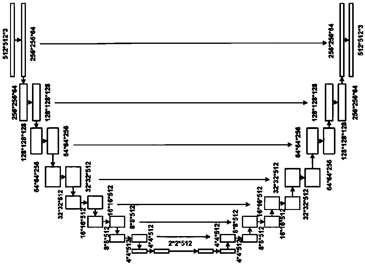
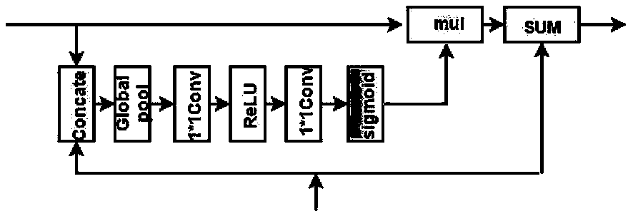
[0023] This method mainly includes three steps: data acquisition and preprocessing, network structure improvement, and model training and testing.
[0024] 1) Data acquisition and preprocessing
[0025] The experimental data set is composed of large-field three-dimensional OCT images collected by a Topcon DRI-OCT scanner with a central wavelength of 1050 nm, and the scanning range includes the center of the macula and the optic nerve head (ONH) area. The collected horizontal scan images were handed over to professional doctors to mark the upper and lower boundaries of the choroid. After the data was acquired, bilinear interpolation was performed on the OCT image and down-sampled to a size of 51...
PUM
 Abstract
Description
Claims
Application Information
 - R&D
- Intellectual Property
- Life Sciences
- Materials
- Tech Scout
- Unparalleled Data Quality
- Higher Quality Content
- 60% Fewer Hallucinations
Browse by: Latest US Patents, China's latest patents, Technical Efficacy Thesaurus, Application Domain, Technology Topic, Popular Technical Reports.
© 2025 PatSnap. All rights reserved.Legal|Privacy policy|Modern Slavery Act Transparency Statement|Sitemap|About US| Contact US: help@patsnap.com


