Pathological image processing method, system and apparatus, and storage medium

A technology of pathological images and processing methods, applied in the field of image processing, can solve the problems of time and energy consumption, low efficiency, etc., and achieve the effects of ensuring accuracy, stable and reliable judgment results, and improving processing and judgment efficiency

Pending Publication Date: 2019-03-29
清影医疗科技(深圳)有限公司
View PDF7 Cites 13 Cited by
  • Summary
  • Abstract
  • Description
  • Claims
  • Application Information

AI Technical Summary

Problems solved by technology

However, observing pathological images and analyzing images with the naked eye takes a lot of time and energy, and is not only inefficient
Moreover, there are more or less differences in the discrimination between different experts.

Method used


Image

  • Pathological image processing method, system and apparatus, and storage medium
  • Pathological image processing method, system and apparatus, and storage medium
  • Pathological image processing method, system and apparatus, and storage medium

Examples


Embodiment 1

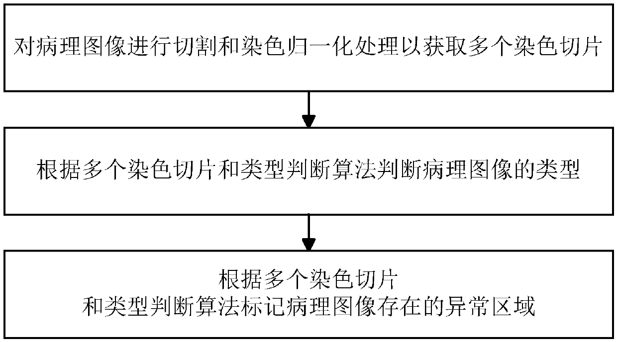
[0037] refer to figure 1 , figure 1 It is a flow chart of a specific embodiment of a pathological image processing method in the present invention, a pathological image processing method, comprising the following steps:

[0038] Image cutting and normalization processing steps, cutting and staining normalization processing on the pathological image to obtain multiple stained slices; the execution order of cutting and staining normalization processing can be reversed, and the pathological image can be cut first to obtain multiple slice, and then perform staining and normalization processing on the slices; it is also possible to firstly carry out staining and normalization processing on the pathological image, and then cut the pathological image after the staining and normalization processing to obtain multiple stained slices. Among them, the pathological image is the region of interest of the histopathological image, that is, the remaining part after removing the background re...

Embodiment 2

[0060] refer to Figure 4 , Figure 4 It is a structural block diagram of a specific embodiment of a pathological image processing system in the present invention, a pathological image processing system, comprising:

[0061] An image cutting and normalization processing unit for cutting and staining normalization processing on the pathological image to obtain multiple stained slices;

[0062] A type judging unit for judging the type of the pathological image according to a plurality of stained slices and a type judging algorithm;

[0063] The abnormal region marking unit is used for marking the abnormal region existing in the pathological image according to the plurality of stained slices and the type judgment algorithm.

[0064] Wherein, in the image cutting and normalization processing unit, the execution order of cutting and staining normalization processing can be reversed, the pathological image can be cut first to obtain multiple slices, and then staining and normaliza...

Embodiment 3

[0067] A pathological image processing device, comprising:

[0068] at least one processor; and,

[0069] a memory communicatively coupled to the at least one processor; wherein,

[0070] The memory stores instructions executable by the at least one processor, and the instructions are executed by the at least one processor, so that the at least one processor can execute the pathological image processing method. For a specific description of the pathological image processing method, refer to the description of Embodiment 1, and details are not repeated here.


PUM


Abstract

The invention discloses a pathological image processing method, system and apparatus, and a storage medium. According to multiple staining slices and type judging algorithm of pathological image to realize automatic judging the type of pathological image, As that technical problem that a large amount of time and energy are required to analyze the pathological image through manual observation, theefficiency is very low and individual discrimination differences exist in the prior art are overcome, the efficiency of processing and judging the pathological image is effectively improved, and the judgement result is stable and reliable; In addition, in order to avoid the influence of image color differences caused by different staining methods, tissue slices at different times, different hospitals or different regions, the pathological images were normalized to ensure the veracity of pathological image type judgement.

Description

technical field [0001] The invention relates to the field of image processing, in particular to a pathological image processing method, system, equipment, and storage medium. Background technique [0002] In the prior art, pathologists analyze the development degree of the disease (normal, inflammation, tumor) by observing information such as cell structure and arrangement in histopathological images. However, observing and analyzing pathological images with the naked eye takes a lot of time and energy, and is not only inefficient. Moreover, the discrimination between different experts also has more or less differences. Contents of the invention [0003] The present invention aims to solve one of the technical problems in the related art at least to a certain extent. Therefore, an object of the present invention is to provide a pathological image processing method, system, device, and storage medium for judging the type of pathological images so as to improve the efficie...

Claims


Application Information

Patent Timeline
Patent Type & Authority Applications(China)
IPC IPC(8): G06T7/00G06T7/10G06K9/62G06K9/46
CPCG06T7/0012G06T7/10G06T2207/30004G06V10/40G06F18/24
Inventor 邹昊郭玉成
Owner 清影医疗科技(深圳)有限公司
Why Eureka
Try Eureka