A cell counting method based on skeleton extraction

A technology for cell counting and skeleton extraction, applied in the field of medical image processing, can solve problems such as separation, impact on histopathological microscopic image classification, cell counting errors, etc., achieve small calculation, good image segmentation effect, and high operating efficiency Effect

Active Publication Date: 2019-04-26
NORTHEASTERN UNIV
View PDF4 Cites 7 Cited by
  • Summary
  • Abstract
  • Description
  • Claims
  • Application Information

AI Technical Summary

Problems solved by technology

[0005] Due to the high adhesion of many tissue cells in histopathological images, the "open" operation cannot completely separate them, resulting in wrong cell counts, which in turn affects the classification of histopathological microscopic images

Method used


Image

  • A cell counting method based on skeleton extraction
  • A cell counting method based on skeleton extraction
  • A cell counting method based on skeleton extraction

Examples


Embodiment Construction

[0034] In order to better explain the present invention and facilitate understanding, the present invention will be described in detail below through specific embodiments in conjunction with the accompanying drawings.

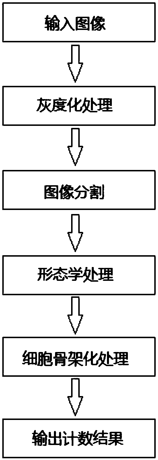
[0035] Such as figure 1 As shown, the present invention provides a method for counting cells based on skeleton extraction, which specifically includes the following steps:

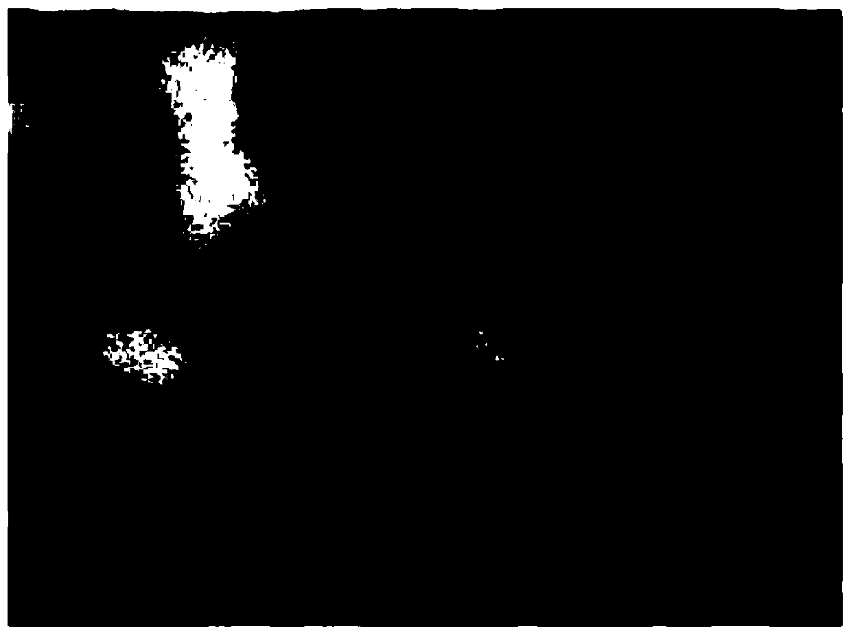
[0036] Step S1. Obtain the histopathological images to be processed, perform grayscale processing on each image to be processed, and then perform image segmentation on each grayscaled image to obtain the Cell binary image.

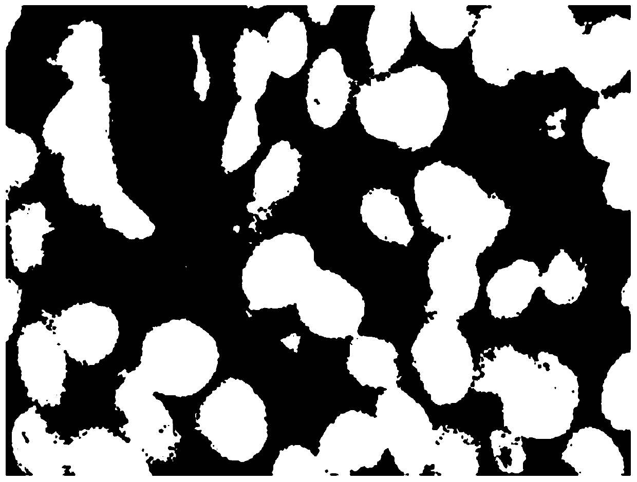
[0037] Step S2, performing morphological processing on each cell binary image to obtain a cell binary image in which intracellular holes are filled, cell edge noise and impurity noise are removed.

[0038] Step S3. Perform cytoskeletal processing on each morphologically processed binary image of cells to obtain the number of cytoskeletal nodes in each image. According...


PUM


Abstract

The invention relates to the technical field of medical image processing and particularly relates to a cell counting method based on skeleton extraction, which comprises the following steps: S1, obtaining a to-be-processed histopathology image, carrying out graying processing on each to-be-processed image, and then carrying out image segmentation on each grayed image to obtain a cell binary imageof each to-be-processed image; S2, performing morphological processing on each cell binary image to obtain a cell binary image with intracellular hole filling and cell edge noise and impurity noise point removal; And S3, performing cell skeletonization processing on each morphologically processed cell binary image to obtain the number of cell skeletonization nodes in each image, and obtaining a cell counting result of each to-be-processed image according to the relationship between the number of cell skeletonization nodes in each image and the number of cells. The obtained cell counting resultis good in accuracy, and the method is high in operation efficiency.

Description

technical field [0001] The invention relates to the technical field of medical image processing, in particular to a method for counting cells based on skeleton extraction. Background technique [0002] Cervical cancer is the most common gynecological malignancy. Although the census of this disease has gradually received attention in recent years, few people go to the hospital for regular checkups in the vast rural areas. There is also a trend of getting younger and younger. Therefore, the histopathological research on cervical cancer is imperative and cannot be delayed. [0003] For a long time, the gap in the level of doctors has been an unavoidable problem. Similarly, the gap in medical level between urban and rural areas still exists. Therefore, it is not difficult to imagine that doctors in some remote areas with backward medical standards or some young and inexperienced doctors must be unable to easily make a judgment on a case. In addition, a doctor has to see more...

Claims


Application Information

Patent Timeline
Patent Type & Authority Applications(China)
IPC IPC(8): G06T7/00G06T7/11G06T7/136G06T7/155G06T7/194G06T5/00
CPCG06T5/002G06T7/0012G06T2207/10056G06T2207/30096G06T2207/30242G06T7/11G06T7/136G06T7/155G06T7/194
Inventor 李晨晏宝恩成奕霖薛丹许宁
Owner NORTHEASTERN UNIV
Why Eureka
Try Eureka