Conductive infusion set guided by central vein headend positioning electrocardio
An infusion set and vein technology, applied in the field of infusion sets, can solve the problems affecting the accuracy of precise positioning of the catheter, and achieve the effects of stable P wave amplitude, high stability and improved accuracy
- Summary
- Abstract
- Description
- Claims
- Application Information
AI Technical Summary
Problems solved by technology
Method used
Image
Examples
Embodiment 1
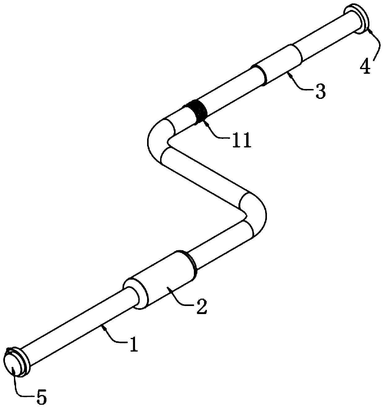
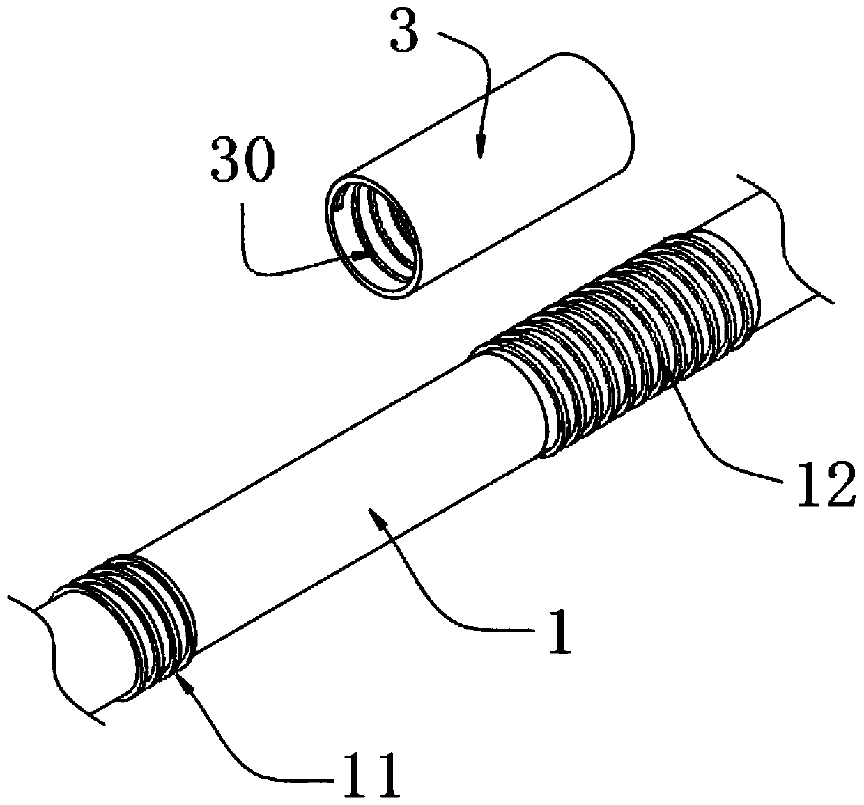
[0027] A central venous head positioning electrocardiographic guidance can be conducted infusion set, such as figure 1 and figure 2 As shown, the infusion tube 1 is included, the end of the infusion tube 1 is provided with a vent hole 10, the dropper 2 is installed on the infusion tube 1, and the position of the infusion tube 1 close to the other end is provided with a protective sleeve 3, inside the protective sleeve 3 An internal thread 30 is provided, a first external thread 11 is provided on the infusion tube 1 on one side of the protective sleeve 3, a second external thread 12 is provided at a position corresponding to the protective cover 3 on the infusion tube 1, and the other end of the infusion tube 1 is installed With luer connector4.
[0028] In this embodiment, the dropper 2 is connected with the infusion tube 1, and the dropper 2 and the infusion tube 1 adopt an integrated structure to ensure the tightness of the connection between the infusion tube 1 and the dr...
Embodiment 2
[0034] As a second embodiment of the present invention, in order to prevent the thickness of the connecting wire 6 from directly affecting the clarity of the P wave amplitude of the electrocardiogram and the accuracy of the precise positioning of the catheter, the inventor made improvements to the infusion tube 1 as a preferred Examples such as Figure 3-5 As shown, the infusion tube 1 corresponding to the second external thread 12 is made of stainless steel.
[0035] In this embodiment, the stainless steel has bright color and good corrosion resistance, which is beneficial to increase the service life.
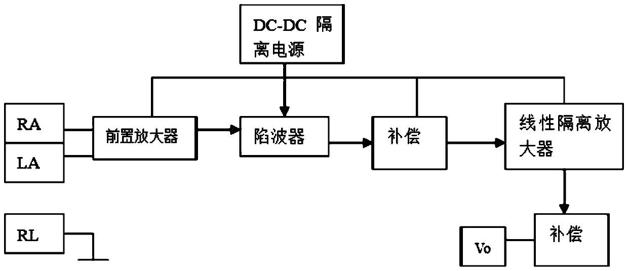
[0036] Furthermore, since human tissue is a volume conductor, ECG signals can be transmitted to all parts of the body. Not only the electrical signals of the heart itself can be measured by cardiac electrodes, but also can be measured by body surface electrodes. Electrodes RA and LA are the main extraction of ECG signals. The point is also the signal source of the intracavit...
Embodiment 3
[0041] As a third embodiment of the present invention, in order to facilitate the use of medical personnel, the end of the inventor's infusion tube 1 is improved. As a preferred embodiment, the vent hole 10 is equipped with a hole cap 5, and the hole cap 5 is connected with the A connection line 6 is provided between the infusion tubes 1 .
[0042] In this embodiment, the hole cap 5 and the connecting wire 6 adopt an integrated structure to ensure the stability of the connection between the hole cap 5 and the connecting wire 6 .
[0043] Specifically, the hole cap 5 is plugged and matched with the vent hole 10. When the vent hole 10 needs to be opened, the hole cap 5 can be separated from the vent hole 10 to realize that the vent hole 10 is opened. When closing, the hole cap 5 and get final product embedded in the air vent 10.
[0044] When the central venous head end positioning electrocardiogram guiding transmissible infusion set of the present invention is in use, through ...
PUM
 Abstract
Description
Claims
Application Information
 - R&D
- Intellectual Property
- Life Sciences
- Materials
- Tech Scout
- Unparalleled Data Quality
- Higher Quality Content
- 60% Fewer Hallucinations
Browse by: Latest US Patents, China's latest patents, Technical Efficacy Thesaurus, Application Domain, Technology Topic, Popular Technical Reports.
© 2025 PatSnap. All rights reserved.Legal|Privacy policy|Modern Slavery Act Transparency Statement|Sitemap|About US| Contact US: help@patsnap.com


