
Image intensity standardization method for brain FLAIR nuclear magnetic resonance image

A technology of nuclear magnetic resonance and image intensity, applied in image enhancement, image data processing, instruments, etc., can solve problems such as lack of image intensity, difference between image intensity and contrast, and influence on the accuracy of segmentation results, and achieve the effect of simple standardization steps

Active Publication Date: 2019-06-07
DALIAN UNIVERSITY
View PDF7 Cites 1 Cited by
  • Summary
  • Abstract
  • Description
  • Claims
  • Application Information

AI Technical Summary

Problems solved by technology

However, the premise of automatic segmentation is that the image has a uniform intensity distribution range and consistent image contrast, otherwise it will affect the accuracy of subsequent segmentation results
Currently, one of the major shortcomings of Magnetic Resonance Image (MRI) is the lack of a standard and quantifiable interpretation of image intensities
Specifically, when using different MRI scanning equipment or different scanning protocols, there are differences in the intensity distribution range and image contrast of the MRI images of different patients
In addition, due to some uncontrollable factors, even if the same MRI equipment is used to scan the same part of the same patient, the scan results at different times still have differences in image intensity and contrast

Method used


Image

  • Image intensity standardization method for brain FLAIR nuclear magnetic resonance image
  • Image intensity standardization method for brain FLAIR nuclear magnetic resonance image
  • Image intensity standardization method for brain FLAIR nuclear magnetic resonance image

Examples


Embodiment 1

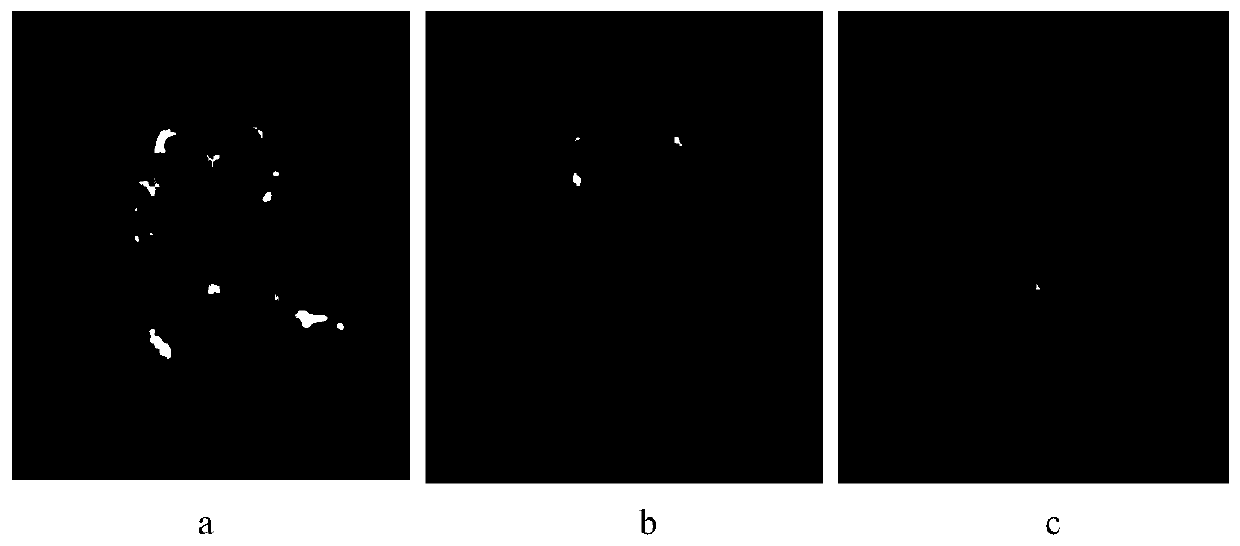
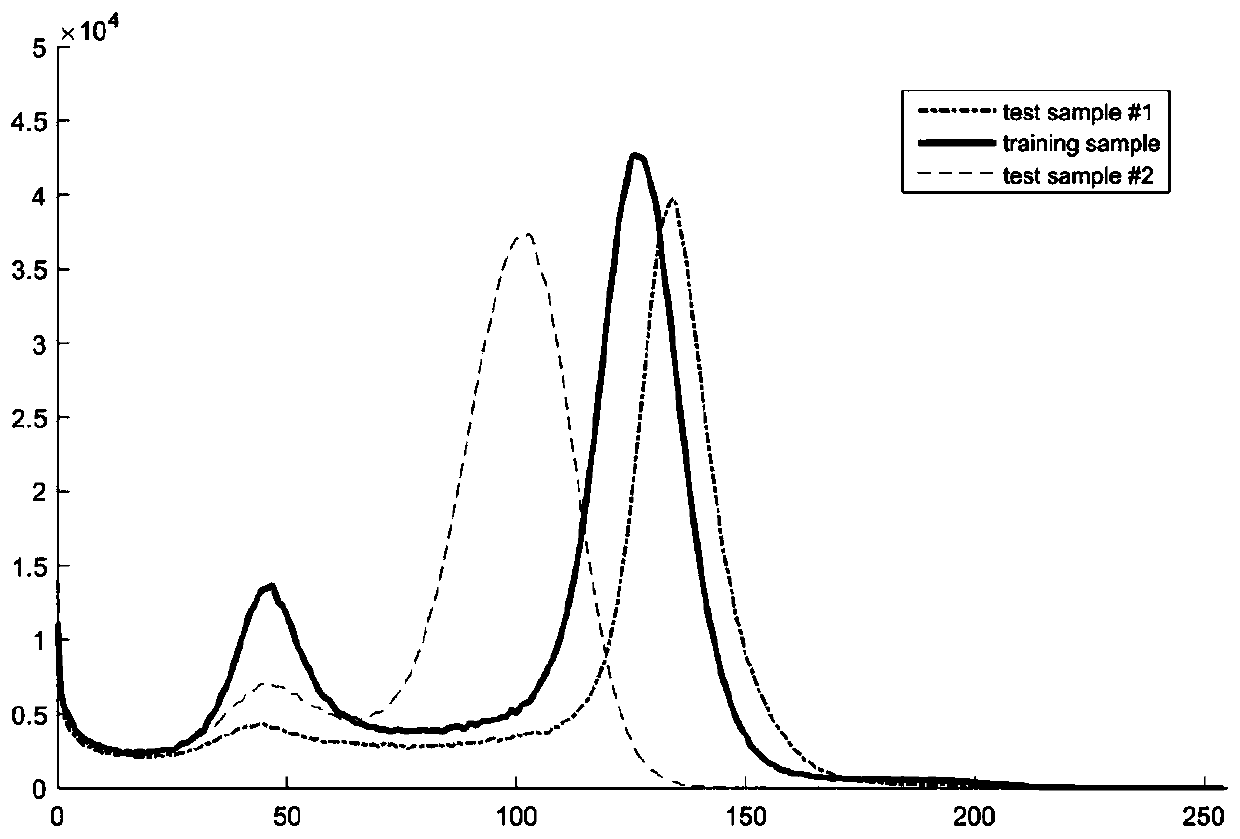
[0050] Two samples are randomly selected from the test sample set as test samples. figure 2 are the slice images located at cross-section 120 / 256 respectively selected from the MRI data of the training sample, test sample #1 and test sample #2. image 3 is the histogram curve of three samples, from image 3 It can be seen that the intensity value corresponding to the peak of test sample 2 is significantly lower than that of the training sample, and the intensity value corresponding to the peak of test sample 1 is close to the training sample, which is why figure 2 The reason why the slice brightness of test sample 1 is close to the training sample, and the slice brightness of test sample 2 is darker than the training sample. Next, standardize and unify the image intensity of the three samples.

[0051] The first step: Calibrate the truncation point of the training sample

[0052] Figure 4 is the histogram of training samples after the initial calibration of the truncati...


PUM


Abstract

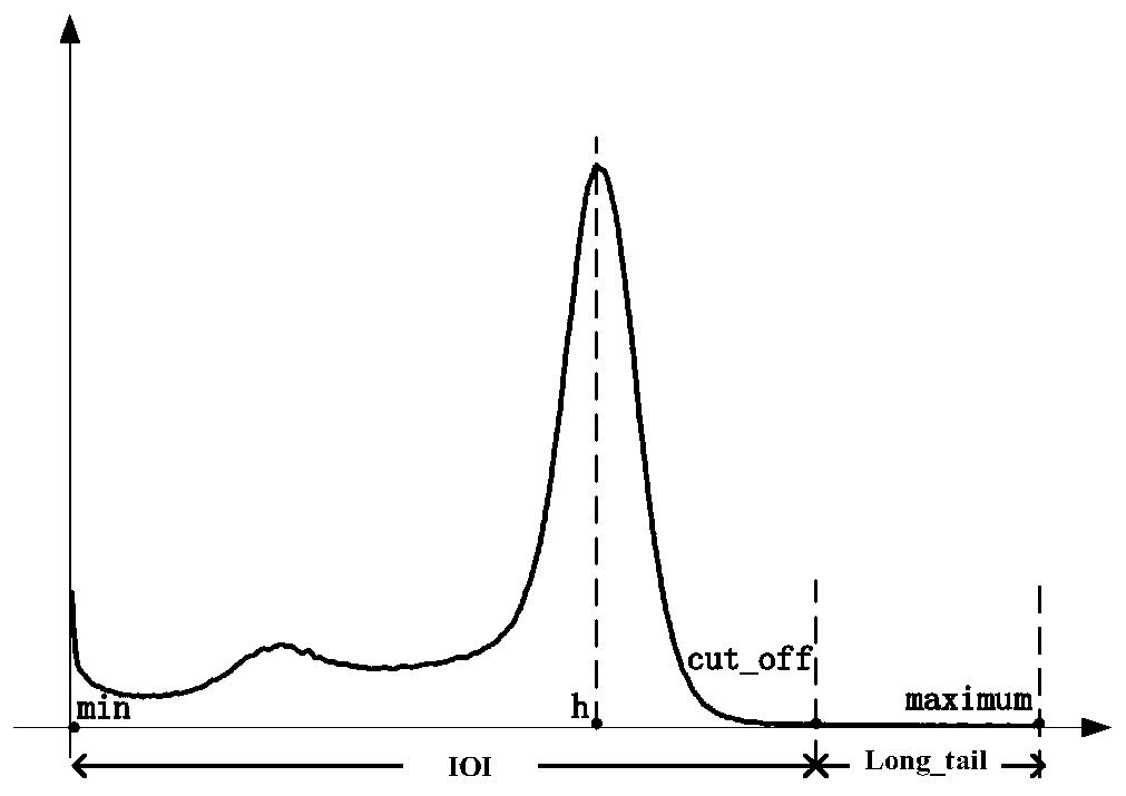
The invention relates to the field of nuclear magnetic resonance image processing, in particular to an image intensity standardization method for a brain FLAIR nuclear magnetic resonance image. The method comprises the following steps: searching a cutoff point on a gray histogram of a FLAIR image, marking voxels with gray values greater than the cutoff point, enabling the voxels not to participatein linear standardization operation, and performing standardization operation on voxels with intensity values less than the cutoff point; through determining a proper truncation point position and carrying out linear normalization, realizing standardization processing of image intensity. According to the method, no matter how many peak values exist in the histogram, only the highest peak value isconcerned, so that the standardization steps are simpler.

Description

technical field [0001] The invention relates to the field of nuclear magnetic resonance image processing, in particular to an image intensity standardization method for brain FLAIR nuclear magnetic resonance images. Background technique [0002] With the development of artificial intelligence technology, automatic segmentation of lesions in brain MRI images has become a research hotspot in brain medical image processing. However, the premise of automatic segmentation is that the image has a uniform intensity distribution range and consistent image contrast, otherwise it will affect the accuracy of subsequent segmentation results. Currently, one of the major shortcomings of Magnetic Resonance Image (MRI) is the lack of a standard and quantifiable interpretation of image intensity. Specifically, when different MRI scanning equipment or different scanning protocols are used, there are differences in the intensity distribution range and image contrast of MRI images of different...

Claims


Application Information

Patent Timeline
IPC IPC(8): G06T5/40
Inventor 赵欣王欣杨晓王洪凯
Owner DALIAN UNIVERSITY
Learn More