Travelling-wave nuclear magnetic resonance method

a nuclear magnetic resonance and travel-wave technology, applied in the field of nuclear magnetic resonance imaging and/or spectroscopy, can solve the problems of increasing static magnetic field strength, reaching their operating limits, and poor application prospects, and achieves less efficiency, simplified safety validation, and more stable electrical performance of remote probes

Inactive Publication Date: 2011-05-19
UNIV ZURICH +1
View PDF2 Cites 69 Cited by
  • Summary
  • Abstract
  • Description
  • Claims
  • Application Information

AI Technical Summary

Benefits of technology

[0049]A setup that generates linearly polarized waves is less efficient in either mode of operation. However it will not require any switching between transmit and receive operation because a linearly polarized wave contains circularly polarized components of both senses of rotation in equal proportion.
[0050]Compared with conventional close-coupling probes, excitation with a remotely placed transmitter has several further advantages. O...

Problems solved by technology

However, an increase in static magnetic field strength requires a concomitant increase in operating radiofrequency.
Moreover, as such RF probes are targeted to work with the sample in their near field, they reach their operating limits and have bad prospects for application if one were to go to even higher field strengths.
In practice, the problems arising when the resonant structures presently used for nuclear magnetic resonance (henceforth: NMR) signal probing are taken to high field strengths are a loss of efficiency and—more detrimentally—a spatially inhomogeneous coupling and a reduction of the size of the volume that can be imaged.
Most of these problems arise due to interference effects of the RF electromagnetic field emitted and/or received by the probe.
For instance, constructive interference yields a signal overshoot in the center region of brain scan...

Method used


Image

  • Travelling-wave nuclear magnetic resonance method
  • Travelling-wave nuclear magnetic resonance method
  • Travelling-wave nuclear magnetic resonance method

Examples


example 1

Travelling Wave MR on a Whole Body System

Theory

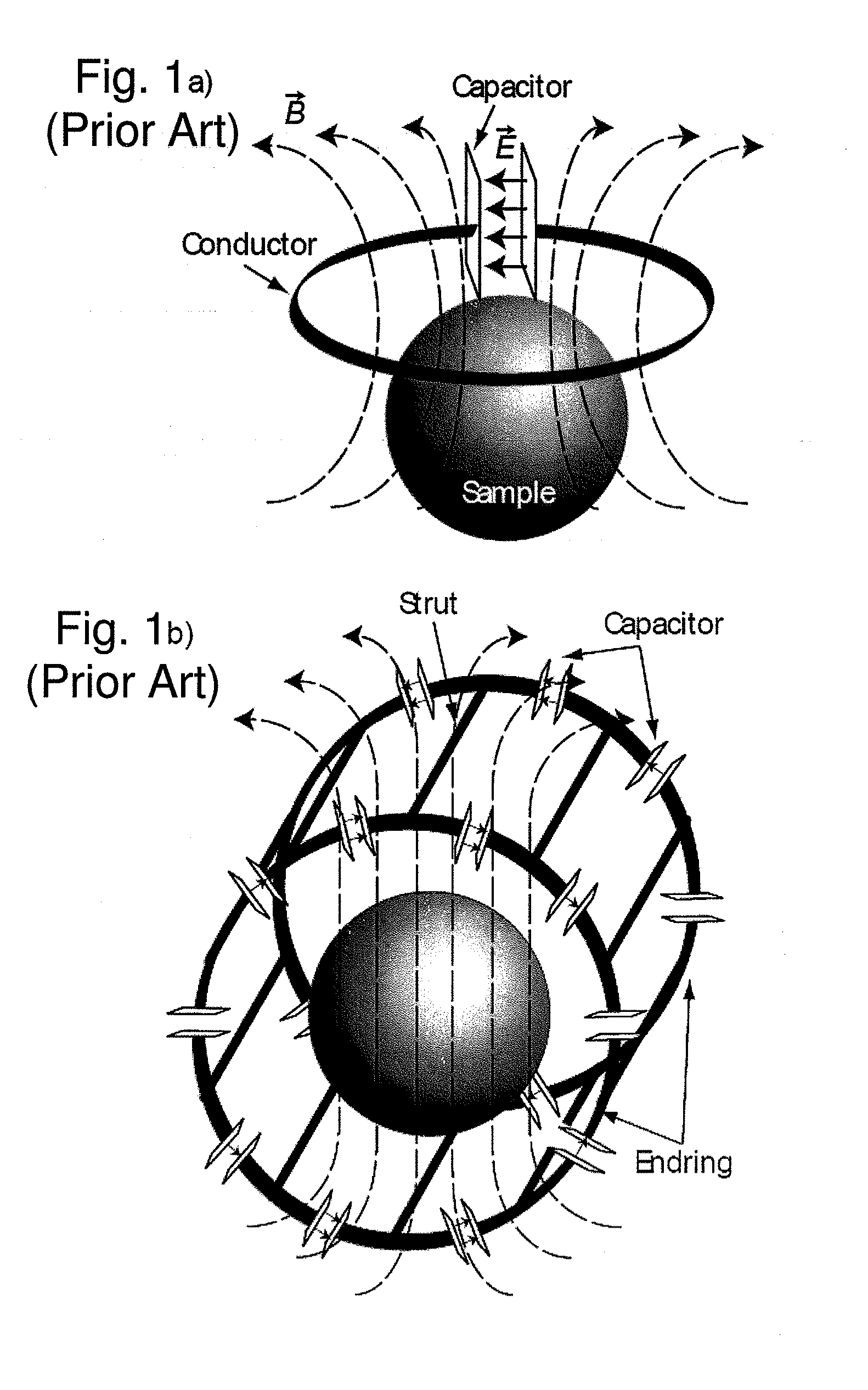
[0081]A propagating plane wave in free space would have maximum homogeneity and transverse polarization down to arbitrarily small wavelengths. Whereas a freely propagating wave cannot be established inside the bore of an NMR magnet, a travelling wave can still be guided by the bore and its RF shield. The modes of the empty bore of typical whole-body 7 Tesla systems (see FIG. 5a) have cutoff frequencies slightly above the proton Larmor frequency. Therefore filling the bore with a preferably lossless dielectric material (in addition to the subject or sample, FIG. 5c) can reduce the cutoff frequencies of the first modes sufficiently ([2]) to support wave propagation at the proton MR frequency.

[0082]In order to prevent axial standing waves stemming from reflections due to guided mode mismatches at the dielectric interfaces of the subject, a di-electric load tapering towards the subject can be placed at the center of the bore (FIG. 5d).

[0083...

example 2

Phantom Experiments

[0089]Further phantom experiments using bottles filled with mineral oil are shown in FIG. 8. The field of view of the images is 530 mm in z direction. The slice of FIG. 8a) was taken while the bore was able to support a travelling wave with minor reflections at both ends of the sample. This was achieved by additional oil bottles of the same diameter placed in the axis of the bore matching the impedance of the modes in the bore to the ones in the sample-filled bore. For comparison the same slice was taken without the matching oil bottles present, see FIG. 8 b). The longitudinal profile taken from both slices depicted in FIG. 8d) show clearly the enhanced homogeneity in axial direction while the transverse field distribution stays unchanged, as shown in FIG. 8c).

example 3

In Vivo Imaging of Feet

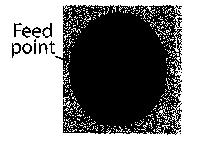
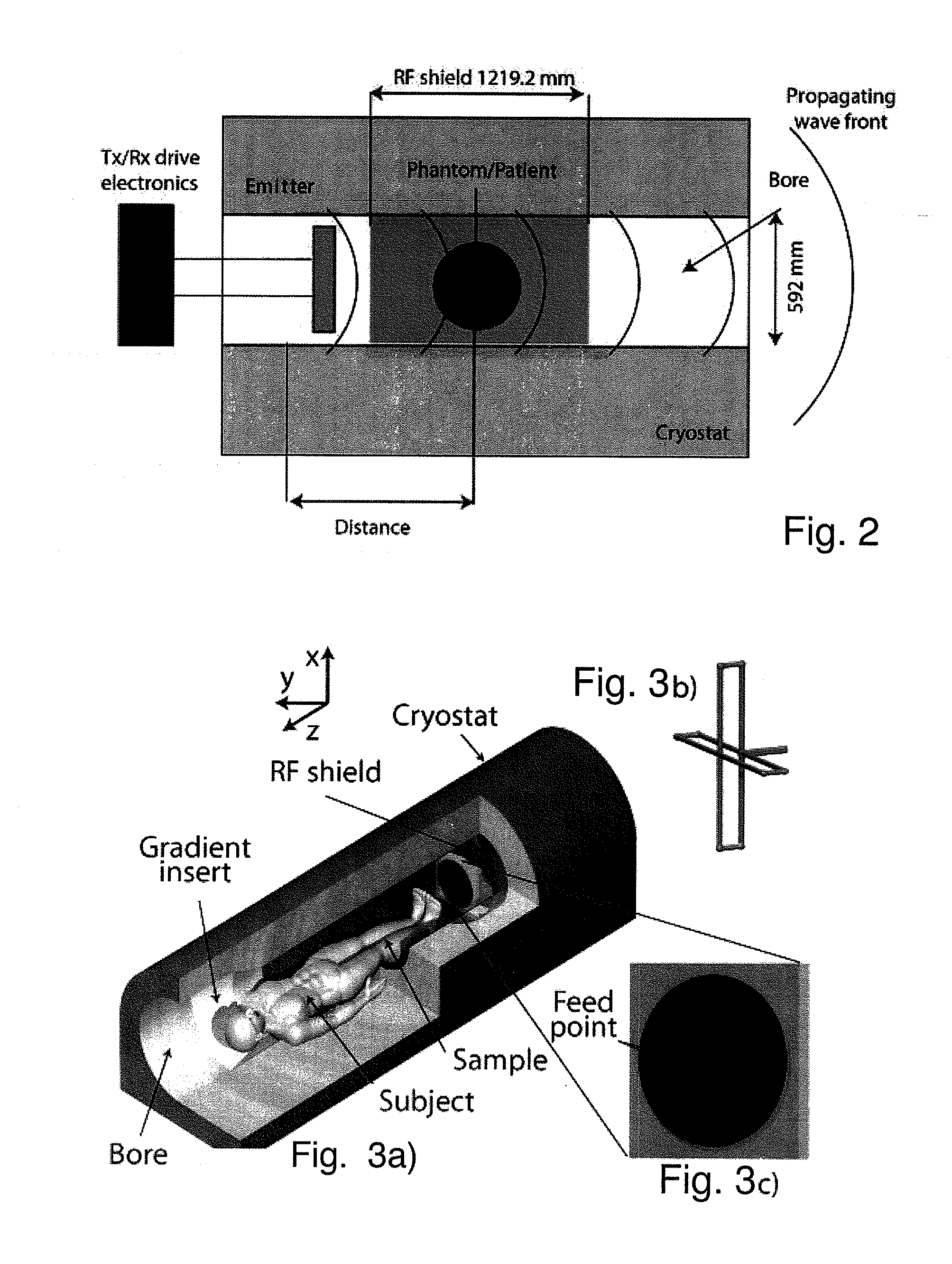
[0090]An example of an in vivo application is given in FIG. 9, which demonstrates MR imaging of a foot of a healthy human subject. The image of FIG. 9 a), which was taken in the travelling wave mode, has comparable quality to the reference image shown in FIG. 9b), which was taken with a conventional 3 T device (birdcage-type body resonator operating at 128 MHz, i.e. the proton MR frequency at 3 T) in near-field mode using the same sequence timings and parameters. However, the field of view in the travelling wave mode was found to be substantially larger and was only limited by the range of the encoding gradient fields. Moreover, an improvement in SNR as compared to the near field mode was found for the travelling wave mode.

Parallel Travelling Waves

[0091]In the examples described so far, a parallel imaging concept was implemented by using a patch antenna with two ports to excite the two degenerate TE11 modes of a circular waveguide, which are the only modes bel...


PUM


Abstract

A method for acquiring an image or spectrum of a subject or object residing within the magnetic field of a magnetic resonance apparatus, comprises the steps of:
    • executing a predetermined pulse sequence for applying gradient magnetic fields and for coupling in electromagnetic excitation pulses to induce nuclear magnetic resonance within the subject or object;
    • detecting an electromagnetic signal resulting from said magnetic resonance; and
    • constructing at least one image or magnetic resonance spectrum of said subject or object from said detected electromagnetic signal.
According to the invention, said coupling in of the electromagnetic excitation pulse and/or said detecting of the electromagnetic signal are carried out substantially by means of travelling electromagnetic waves.

Description

CROSS REFERENCE TO RELATED APPLICATIONS[0001]This application claims the benefit of PCT Application No. PCT / EP2009 / 002846, filed on Apr. 17, 2009 and European Patent Application No. 08007601.1, filed Apr. 18, 2008, the disclosures of which are herein incorporated by reference in their entirety.FIELD OF THE INVENTION[0002]The present invention generally relates to nuclear magnetic resonance imaging and / or spectroscopy.BACKGROUND OF THE INVENTION[0003]Nuclear Magnetic Resonance Imaging (henceforth: MRI), in particular for imaging of selected portions of a patient or another object, has so far relied on two main categories of radiofrequency (RF) coils, as generally described in U.S. Pat. No. 4,920,318. The first type is a volume resonator dimensioned to be disposed around the entire object or patient to be imaged or around a portion thereof. The other type of coil is generally formed by wrapping wire or other conductors on a flat dielectric sheet shaped in such way to be positionable a...

Claims


Application Information

Patent Timeline
IPC IPC(8): G01R33/48
CPCG01R33/4802G01R33/345
Inventor FROHLICH, JURGDE ZANCHE, NICOLAPRUSSMANN, KLAASBRUNNER, DAVID
Owner UNIV ZURICH
Why Eureka
Try Eureka