
A method, device and system for generating pictures

A picture and regeneration technology, applied in the medical field, can solve problems such as deformation and distortion, and achieve the effect of improving the accuracy of diagnosis

Active Publication Date: 2022-03-29
VISIONVERA INFORMATION TECH CO LTD
View PDF4 Cites 0 Cited by
  • Summary
  • Abstract
  • Description
  • Claims
  • Application Information

AI Technical Summary

Problems solved by technology

[0005] The method, device and system of a method for generating pictures provided by the present invention solve the cutting technology used in generating pictures. When the edge is insufficient to preset pixels, it is still cut according to the preset pixels, so that the picture is stretched, resulting in deformation and distortion The problem

Method used


Image

  • A method, device and system for generating pictures
  • A method, device and system for generating pictures
  • A method, device and system for generating pictures

Examples


Embodiment Construction

[0056] The following will clearly and completely describe the technical solutions in the embodiments of the present invention with reference to the accompanying drawings in the embodiments of the present invention. Obviously, the described embodiments are some of the embodiments of the present invention, but not all of them. Based on the embodiments of the present invention, all other embodiments obtained by persons of ordinary skill in the art without creative efforts fall within the protection scope of the present invention.

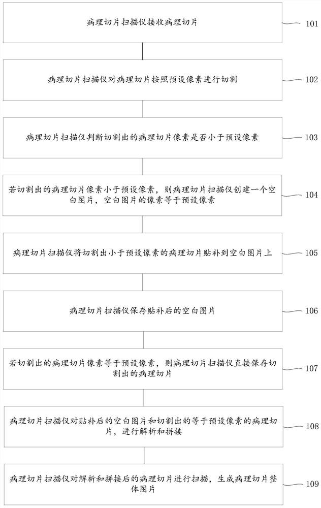
[0057] figure 1 A flow chart of a method for generating pictures according to an embodiment of the present invention is shown. The method is applied in the medical field, and the medical field includes: a pathological slice scanner, which is an intelligent device for digitizing pathological slices and generating pictures. The method includes the following steps:

[0058] Step 101: the pathological slice scanner receives the pathological slice.

[005...


PUM


Abstract

The invention provides a method, device and system for generating pictures. The method is applied in the medical field, and the medical field includes: a pathological slice scanner, and the method includes: the pathological slice scanner receives the pathological slice, cuts the pathological slice according to preset pixels, and judges whether the cut out pathological slice pixels are If it is smaller than the preset pixel, a blank image will be created, and the cut pathological slice smaller than the preset pixel will be patched onto the blank image, and the patched blank image will be saved. If it is equal to the preset pixel, the cut out image will be saved directly. The pathological slices of the patched blank images and the pathological slices equal to the preset pixels are analyzed and stitched together, and then scanned to generate the overall picture of the pathological slices. According to the invention, the overall picture of the generated pathological slice is complete and clear, and the edge area will not be stretched, and will not be deformed and distorted, thereby improving the accuracy of diagnosis of diseases by medical workers.

Description

technical field [0001] The present invention relates to the medical field, in particular to a method, device and system for generating pictures. Background technique [0002] Pathological sectioning is a medical method widely used in the medical field. Take a certain size of diseased tissue and make pathological sections with histopathological methods (usually the diseased tissue is embedded in paraffin block, cut into thin slices with a microtome, and then hematoxylin Essence - eosin (H-E) staining), further examination of the occurrence and development of the lesion with a microscope, and finally a pathological diagnosis. It is a kind of pathological specimen. During the production, some diseased tissues or organs are treated with various chemicals and embedding methods to make them fixed and hardened, cut into thin slices on a microtome, adhered to glass slides, and stained with Various colors for inspection under a microscope to observe pathological changes, make pathol...

Claims


Application Information

Patent Timeline
Patent Type & Authority Patents(China)
IPC IPC(8): G16H30/40
CPCG16H30/40
Inventor 谢文龙吕亚亚李云鹏沈军
Owner VISIONVERA INFORMATION TECH CO LTD
Learn More