Fundus image focus area labeling method based on deep learning

A fundus image and deep learning technology, applied in the field of medical image processing, can solve the problems of high price and small scale, and achieve the effect of improving the generation quality, reducing the probability of misdiagnosis and missed diagnosis, and reducing the cost of manual labeling.

Pending Publication Date: 2019-08-06
南京星程智能科技有限公司
View PDF3 Cites 30 Cited by
  • Summary
  • Abstract
  • Description
  • Claims
  • Application Information

AI Technical Summary

Problems solved by technology

However, medical annotation images are usually small in scale and expensive to annotate, so we hope to use a simple and direct way to directly generate the annotations of the lesion area from the fundus retinal image to reduce the cost of manual annotation

Method used


Image

  • Fundus image focus area labeling method based on deep learning
  • Fundus image focus area labeling method based on deep learning
  • Fundus image focus area labeling method based on deep learning

Examples


Embodiment 1

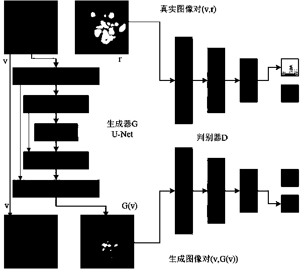
[0026] Implementation example 1: The deep learning-based fundus image lesion area labeling method provided by the present invention automatically labels the hard exudate lesion area in the original image of diabetic retinopathy, refer to figure 1 As shown, the fundus image lesion area labeling method based on deep learning mainly includes the following steps:

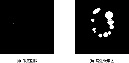
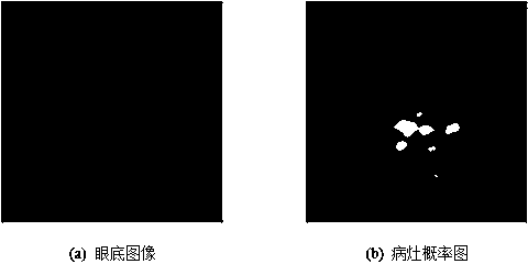
[0027] Step 1: Select a dataset. The data set used in the present invention is the DIARETDB1 data set, wherein the DIARETDB1 data set is a color fundus image collected by Kuopio University Hospital for DR detection, including 89 color fundus images, 48 ​​of which contain hard exudates 41 images without hard exudates, and each image has a size of 1152×1500. When the images that do not contain hard exudates are also used for training, the lesion probability map obtained in the experiment cannot get the expected results, and the output is a completely black image, so only 48 images containing hard exudates are selected. ...


PUM


Abstract

The invention discloses a fundus image focus area labeling method based on deep learning, and the method comprises the steps: selecting a sample, and carrying out the preprocessing: carrying out the cutting of an image, and carrying out the horizontal and vertical overturning and normalization processing; constructing a convolutional neural network and a deconvolutional neural network as an imagegenerator, inputting the preprocessed color fundus image, and outputting a corresponding focus probability graph; constructing a convolutional neural network as a discriminator, inputting and generating a focus image and a real focus image, and outputting a probability that the focus image is judged to be a real image; alternately training the generation network and the discrimination network until a satisfaction result can be generated; and marking a focus area in the fundus image according to the generated focus probability graph. The fundus image focus probability graph is generated by using the deep convolutional neural network, and the fundus image focus area is automatically labeled. The automatic labeling can provide an auxiliary basis for the diagnosis of doctors, and meanwhile, the cost of manual labeling can be greatly reduced.

Description

technical field [0001] The invention relates to a deep learning-based labeling method for fundus image lesion regions, which belongs to the field of medical image processing. Background technique [0002] In recent years, with the continuous improvement of medical imaging acquisition equipment and the continuous development of image processing, pattern recognition, machine learning and other disciplines, the field of multidisciplinary medical image processing and analysis has achieved fruitful results. These achievements are of great significance for assisting doctors in making rapid and accurate diagnoses. [0003] Diabetic retinopathy (Diabetic Retinopathy, DR) is a common complication of diabetes and one of the main causes of blindness in middle-aged and elderly people. thing. The rate of blindness can be effectively reduced through regular screening and early diagnosis of diseases. Due to the huge number of people who need to be screened, it is a time-consuming and la...

Claims


Application Information

Patent Timeline
Patent Type & Authority Applications(China)
IPC IPC(8): G06T7/11G06T7/13G06T7/136G06N3/04G06K9/62
CPCG06T7/11G06T7/13G06T7/136G06T2207/20081G06T2207/20084G06T2207/30041G06N3/048G06N3/045G06F18/214
Inventor 万程俞秋丽周鹏彭琦吴陆辉华骁
Owner 南京星程智能科技有限公司
Why Eureka
Try Eureka