
Pathological image multi-staining separation method based on deep learning

A technology of pathological images and deep learning, applied in instruments, character and pattern recognition, computer components, etc., can solve problems such as insufficient deconvolution images, achieve good generalization ability, and improve the effect of dyeing separation performance

Active Publication Date: 2019-08-09
NANTONG UNIVERSITY
View PDF4 Cites 5 Cited by
  • Summary
  • Abstract
  • Description
  • Claims
  • Application Information

AI Technical Summary

Problems solved by technology

They show that while NMF performs better, neither method is sufficient to fully deconvolute the image

Method used


Image

  • Pathological image multi-staining separation method based on deep learning
  • Pathological image multi-staining separation method based on deep learning
  • Pathological image multi-staining separation method based on deep learning

Examples


Embodiment

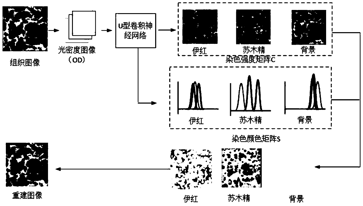
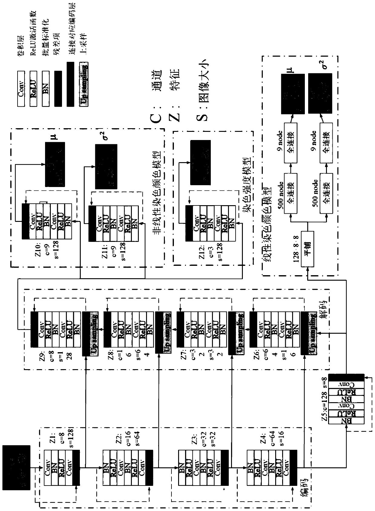
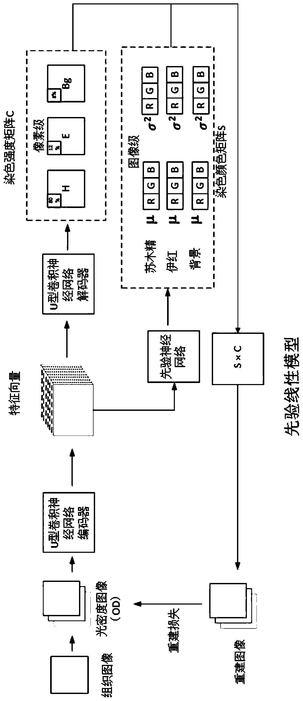
[0069] The raw images were densitometrically transformed and input into a ResU-Net model with a 12-level architecture for multi-task staining separation. The ResU-Net model is supported by three parts: contraction path, bridging path, and expansion path to complete linear staining color matrix prediction, nonlinear staining color matrix prediction and staining concentration prediction. The contraction path consists of the first 1-4 stages of the network, which are used to reduce the spatial dimension of the feature map, and at the same time increase the number of feature maps layer by layer, and extract the input image as compact features. Level 5 is the bridging part that connects the contraction and expansion paths and implements the linear dye color matrix prediction function. The expansion path is composed of a 6-9 level network, which is used to gradually restore the details of the target and the corresponding spatial dimensions. The expansion path will be up-sampled, and...


PUM


Abstract

The invention discloses a pathological image multi-staining separation method based on deep learning, and the method comprises the following steps: (1) carrying out the optical density transformationof a pathological staining image, and obtaining an optical density matrix of an original pathological staining image; (2) constructing a ResU-Net model by using the optical density matrix obtained inthe step (1); (3) training the ResU-Net model obtained in the step (2); and (4) carrying out image dyeing separation through the ResU-Net model trained in the step (3). According to the method, pixel-level analysis can be carried out on the original image, and the same kind of tissue in the image is better separated, so that the dyeing separation performance is improved.

Description

technical field [0001] The invention relates to the technical field of image information processing, in particular to a method for multi-staining separation of pathological images based on deep learning. Background technique [0002] Stain separation is a preprocessing technique used to aid in the automated analysis of histopathology images. Chemical reagents that bind to specific structures can enhance the discrimination of different tissue types. In histopathology, the most widely used stain is hematoxylin and eosin (H&E). Hematoxylin binds to nucleic acids, giving cell nuclei a dark blue or purple color, while eosin binds to proteins in tissue, giving the cell matrix a pink color. Traditional staining methods such as color deconvolution (CD) and independent component analysis (ICA) aim to find the optimal staining matrix and staining concentration matrix, while the predefined values ​​of the staining matrix are imprecise. And in practice, the observed staining with hem...

Claims


Application Information

Patent Timeline
IPC IPC(8): G06K9/00G06K9/46
CPCG06V20/698G06V10/56
Inventor 张堃付君红李子杰姜朋朋吴建国张培建陆平
Owner NANTONG UNIVERSITY
Learn More