Analysis system for scanned pathological section image and scanning method thereof

An analysis system, a technology for pathological slices, applied in the direction of analyzing materials, material analysis by optical means, measuring devices, etc., can solve the problems of fast focusing, defocusing, rough and uneven surface of slices, etc.

Active Publication Date: 2019-08-23
广州英特美迪科技有限公司 +1
View PDF6 Cites 7 Cited by
  • Summary
  • Abstract
  • Description
  • Claims
  • Application Information

AI Technical Summary

Problems solved by technology

Most of the existing slide scanning systems use a microscope with a magnification of 20 to 40 times. The depth of field of a microscope with a 20 times magnification is generally between 1 micron and 1.4 microns depending on the brand. Because the surface of the slice is often rough and not Smooth, so that the near part of the image in the field of view observed by the objective lens is clear, but the far part is blurred. Therefore, in the process of slice scanning, due to the complicated structure of the mechanical focusing method of the microscope, the movement accuracy of the stage is limited by the mechanism, which cannot meet the requirements. The requirement of fast focusing may easily lead to inaccurate focusing and out-of-focus phenomena

Method used


Image

  • Analysis system for scanned pathological section image and scanning method thereof
  • Analysis system for scanned pathological section image and scanning method thereof
  • Analysis system for scanned pathological section image and scanning method thereof

Examples


Embodiment Construction

[0138] The embodiments of the present invention will be described in detail below.

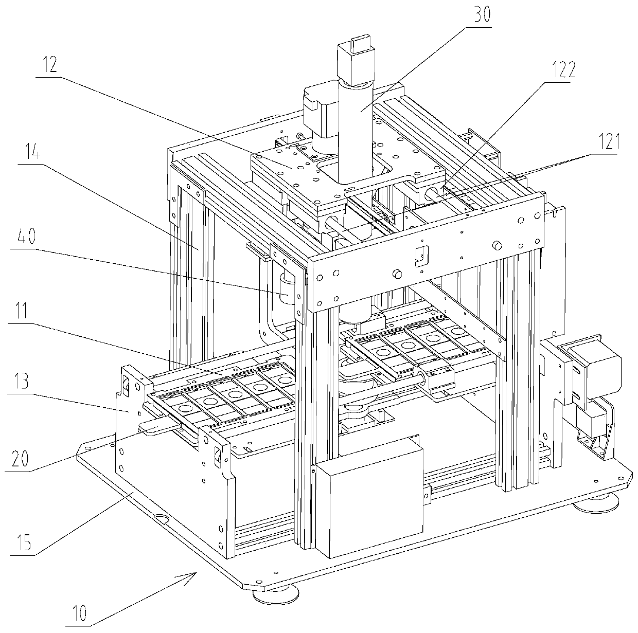
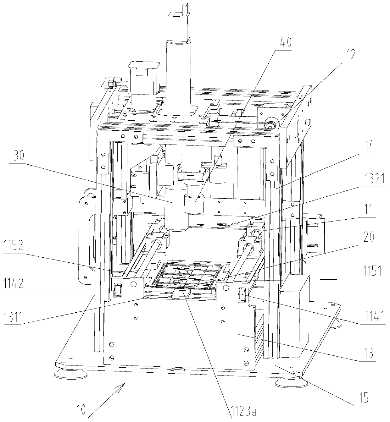
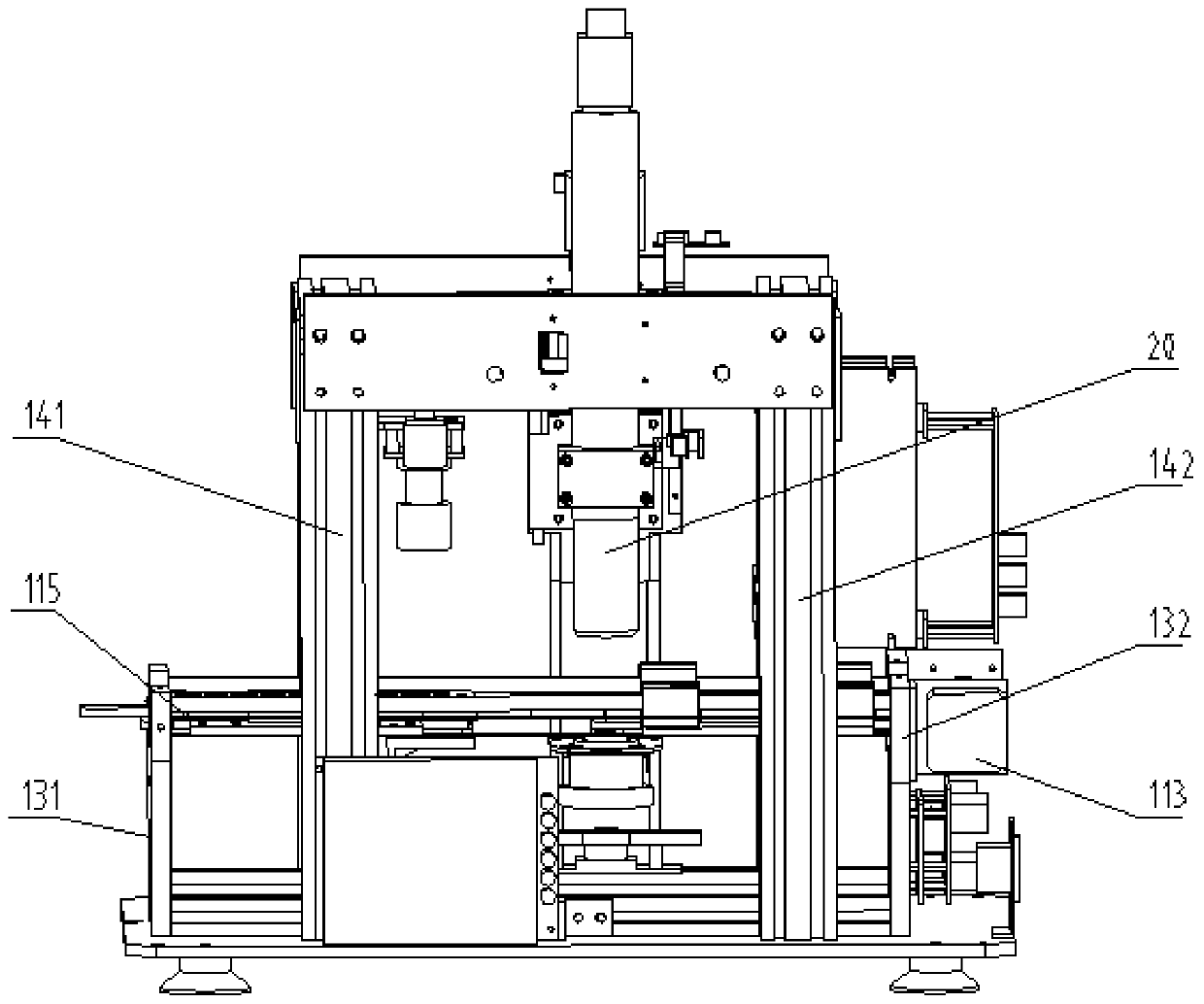
[0139] The scanning method of the pathological section scanning image analysis system is applied to the glass slide imaging scanning mirror 30 of the pathological section scanning image analysis system. The glass slide imaging scanning mirror 30 includes a microscope barrel 31 and a main camera 33, and the scanning method includes:

[0140] Reduce the magnification of the microscope barrel 31, thereby increasing the depth of field length of the microscope barrel 31, and obtain a larger clear imaging range; the magnification of the microscope barrel 31 is six times to fourteen times;

[0141] The imaging pixels of the main camera 33 are increased to obtain a higher pixel density; the imaging pixels of the main camera 33 are greater than or equal to 4.5 million pixels.

[0142] The microscope barrel 31 includes an eyepiece end and an objective lens end, and the main camera 33 is mounted on the e...


PUM


Abstract

The present invention discloses an analysis system for a scanned pathological section image and a scanning method thereof. The scanning method of the analysis system for the scanned pathological section image is applied to a slide camera scanning mirror of the analysis system for the scanned pathological section image. The slide camera scanning mirror comprises a microscope tube and a main camera.The scanning method comprises the following steps: a magnification of the microscope tube is reduced, thereby a field depth of the microscope tube is increased and a larger clear imaging range is obtained; and pixels of the main camera are increased to obtain a higher pixel density. The scanning method of the analysis system for the scanned pathological section image is to increase the field depth of the microscope tube and improve the pixels of the main camera by reducing the magnification of the microscope tube, thereby the pixel density and clarity that only are achieved by using the microscope tube with an original magnification can be achieved. The longer the field depth, the larger the range corresponding to clear imaging, and the greater the tolerance. So the difficulty of focusingis reduced and fast focusing is available. At the same time, the requirement on movement accuracy of a moving part of a mechanical structure is relatively low.

Description

technical field [0001] The invention relates to the field of medical test equipment, in particular to a pathological slice scanning image analysis system and a scanning method thereof. Background technique [0002] At present, in the section scanning process performed by the section scanning system, in order to obtain higher definition, it is generally desirable to use a microscope with a larger magnification. Most of the existing slice scanning systems use microscopes with a magnification of 20 times to 40 times. The depth of field of a microscope with a magnification of 20 times is generally between 1 micron and 1.4 microns depending on the brand. It is smooth, so that the image in the field of view observed by the objective lens is clear near and blurred in the distance. Therefore, in the process of slice scanning, due to the complex structure of the microscope's mechanical focusing method, the moving accuracy of the stage is limited by the mechanism, which cannot meet th...

Claims


Application Information

Patent Timeline
Patent Type & Authority Applications(China)
IPC IPC(8): G01N21/00G01N21/01
CPCG01N21/00G01N21/01G01N2021/0112
Inventor 方陕渊张凯闻
Owner 广州英特美迪科技有限公司
Why Eureka
Try Eureka