Reconstruction method for removing CT cone beam artifacts

A cone-beam and normalization technology, applied in the reconstruction field of removing CT cone-beam artifacts, can solve problems such as uneven intensity and affecting imaging quality, and achieve the effect of improving image quality, improving coherence and efficiency

Pending Publication Date: 2019-09-20
FMI MEDICAL SYST CO LTD
View PDF7 Cites 1 Cited by
  • Summary
  • Abstract
  • Description
  • Claims
  • Application Information

AI Technical Summary

Problems solved by technology

[0004] For reconstruction, the cone beam artifact effect is also more significant, especially in the image of insufficient data area, it is easy to have uneven intensity, which affects the imaging quality, so it is necessary to accurately acquire the data in the insufficient data area. Cone Beam Artifact Correction

Method used


Image

  • Reconstruction method for removing CT cone beam artifacts
  • Reconstruction method for removing CT cone beam artifacts
  • Reconstruction method for removing CT cone beam artifacts

Examples


Embodiment Construction

[0044] In order to make the present invention more obvious and understandable, a preferred embodiment is now described in detail in conjunction with the accompanying drawings.

[0045] A reconstruction method for removing CT cone-beam artifacts of the present invention. The reconstruction method includes a training process and a generating process. The training process refers to a method of pre-training a neural network and is the basis for obtaining a usable neural network; The generation process is to call the neural network method. Once the neural network is trained, the original data can be corrected to complete the reconstruction work.

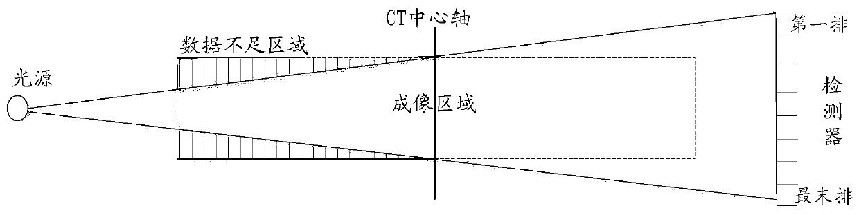
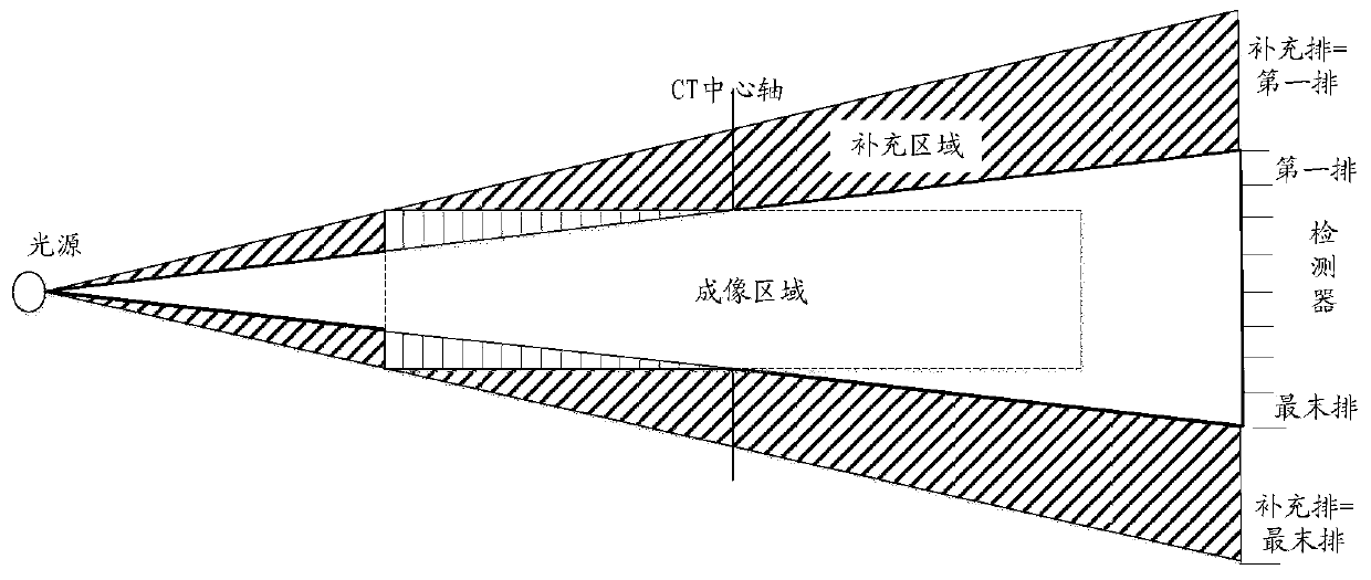
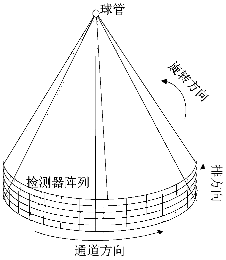
[0046] Such as Figure 4 As shown, the training process is used to calculate the specific weights of neurons in the neural network. The input starting point is the sine data of CT. The specific steps are:

[0047] Step A1. Define the sine data collected by the original CT and corrected as A0, and A0 is the three-dimensional data, which are arran...


PUM


Abstract

The invention provides a reconstruction method for removing CT cone beam artifacts. The reconstruction method comprises: A1, defining original CT data A0; A2, carrying out downsampling to obtain AL; A3, normalizing the data to obtain ALNorm; A4, transmitting the ALNorm to a neural network for training, and generating a training result PA0; B1, defining original CT data B0; B2, performing downsampling to obtain BL; B3, normalizing the data to obtain BLNorm; B4, transmitting the BLNorm into the neural network, and generating an expanded generation result PB1; B5, performing strength recovery on the PB0 according to the inverse operation of data normalization to obtain a PN; B6, performing upsampling processing on the PN according to the inverse operation of downsampling to obtain PU2D; B7, replacing B0 with a corresponding position in the middle of the PU2D to obtain BN; and B8, outputting the result BN and continuing CT reconstruction to obtain an image domain result. The image quality can be improved.

Description

Technical field [0001] The present invention relates to the field of CT imaging technology, in particular to a reconstruction method for removing CT cone beam artifacts. Background technique [0002] CT (Computed Tomography), that is, electronic computer tomography, it uses precisely collimated X-ray beams, gamma rays, ultrasound, etc., together with a very sensitive detector to scan a certain part of the human body one by one. It has the characteristics of fast scanning time and clear image, which can be used for the inspection of various diseases; according to the different radiation used, it can be divided into: X-ray CT (X-CT), ultrasound CT (UCT) and γ-ray CT (γ-CT) )Wait. [0003] In order to achieve faster, more accurate, and low-dose scanning in modern commercial CT, the number of detector rows is constantly increasing. In recent years, CT has entered the medical application field from 16 rows, 64 rows to 256 rows. However, as the number of rows increases, the angle betw...

Claims


Application Information

Patent Timeline
IPC IPC(8): G06T11/00G06K9/62G06N3/04G06T5/00G06T5/50
CPCG06T11/008G06T5/50G06N3/04G06T2207/10081G06F18/214G06T5/70
Inventor 褚政阿泽子·伊赫莱夫王瑶法郭洪斌金燕南
Owner FMI MEDICAL SYST CO LTD
Why Eureka
Try Eureka