Lung tissue image segmentation method based on deep learning

A tissue image, deep learning technology, applied in image analysis, image generation, image enhancement and other directions, can solve the problem of inaccurate segmentation, achieve high segmentation accuracy, solve local convergence and false positive segmentation, and solve the problem of false positive segmentation. Effect

Inactive Publication Date: 2019-10-08
BEIJING JIAOTONG UNIV
View PDF3 Cites 14 Cited by
  • Summary
  • Abstract
  • Description
  • Claims
  • Application Information

AI Technical Summary

Problems solved by technology

However, purely from the visualization results, for images with dissimilar shapes in the database, the

Method used


Image

  • Lung tissue image segmentation method based on deep learning
  • Lung tissue image segmentation method based on deep learning
  • Lung tissue image segmentation method based on deep learning

Examples


Embodiment

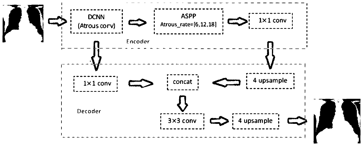
[0038] Embodiment 1 of the present invention provides a lung tissue image segmentation method based on deep learning, including:

[0039] The X-ray chest image is input into the model, wherein the model is obtained by training with multiple sets of training data, and each set of training data in the multiple sets of training data includes: the X-ray chest image and the The gold standard for lung tissue in images;

[0040] Acquiring output information of the model, wherein the output information includes a segmentation result of lung tissue in the X-ray chest film.

[0041] In Example 1 of the present invention, after verification on public datasets and pneumoconiosis datasets, better segmentation performance can be obtained when segmenting chest radiograph tissue than deep learning methods such as SCAN. The realization steps of the present invention are as follows:

[0042] Step 1: Mark the gold standard for the pneumoconiosis data set, and perform data enhancement on the ma...

Embodiment 2

[0052] Embodiment 2 of the present invention provides a deep learning method for X-ray chest film segmentation. After verification on public datasets and pneumoconiosis datasets, this method can obtain better segmentation performance than deep learning methods such as SCAN in segmenting chest radiographs. The realization steps of the present invention are as follows:

[0053] (1) Training part

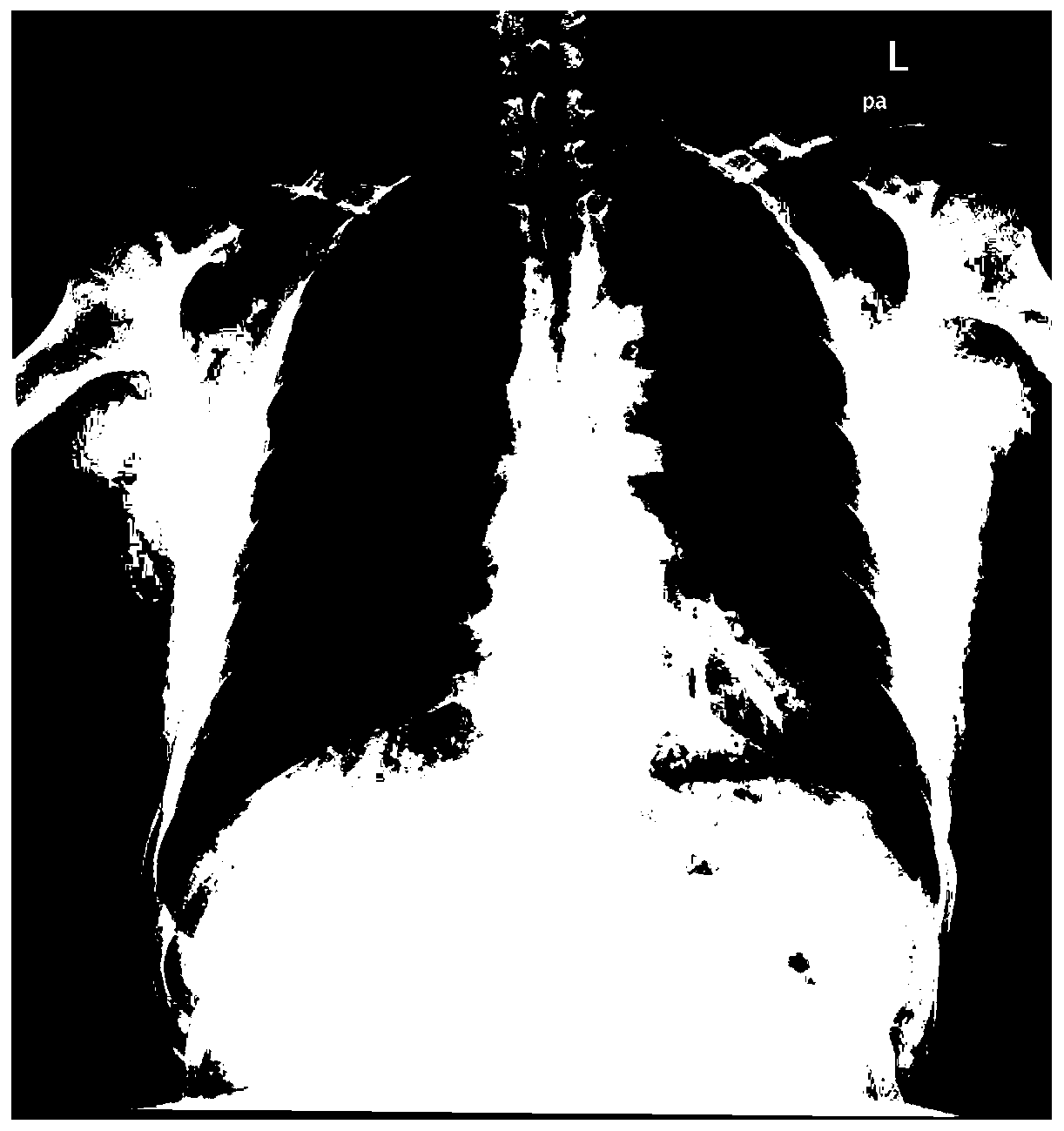
[0054] There are two types of X-ray chest X-ray public datasets with complete labels for lung tissue segmentation: TB dataset and JSRT dataset. The JSRT dataset is 89 pathological images of pulmonary nodules published by the Japanese Radiological Society, such as figure 2 As shown, it is an image case in the JSRT dataset; the TB dataset contains 139 pulmonary tuberculosis pathological images, such as image 3 As shown, it is an example of an image in TB data. Experiments on public datasets use the JSRT dataset as the training set and the TB dataset as the test set.

[0055] Such ...


PUM


Abstract

The invention provides a lung tissue image segmentation method based on deep learning, and belongs to the technical field of medical image segmentation. The lung tissue image segmentation method comprises the steps that an X-ray chest radiograph image is input into a segmentation model, the segmentation model is obtained through training of multiple sets of training data, and each set of trainingdata in the multiple sets of training data comprises the X-ray chest radiograph image and a corresponding gold standard used for identifying lung tissue; and output information of the model is obtained, and the output information comprises a segmentation result of the lung tissue in the X-ray chest radiography image. According to the lung tissue image segmentation method, the segmentation of the lung tissue of the X-ray chest radiography is realized through an improved Deeplabv3+ deep learning method, and the problems of local convergence and false positive segmentation when the lung tissue issegmented by using a traditional method are solved; the lung tissue image segmentation method respectively obtains 95.3% of MIoU and 94.8% of MIoU on the public data set and the pneumoconiosis data set; and the false positive problem of the FCN network is solved, and the segmentation accuracy of ribs at the thoracic diaphragm angle and on the X-ray chest radiography in the SCAN network method isimproved.

Description

technical field [0001] The invention relates to the technical field of medical image segmentation, in particular to a deep learning-based lung tissue image segmentation method that improves the accuracy of lung tissue segmentation and reduces the impact of noise on pneumoconiosis classification and identification. Background technique [0002] With the diversification and maturity of medical imaging technology, medical image research has become one of the hot research fields. Based on the multiplicity and diversity of lung diseases, computer-aided diagnosis has become an important means of clinical treatment. Therefore, high-accuracy technical support is essential. At present, lung tissue segmentation techniques are roughly divided into three categories: traditional methods, traditional machine learning methods, and deep learning methods. The traditional method for segmenting lung tissue has high requirements on the data set, not only the strong boundary information of the ...

Claims


Application Information

Patent Timeline
IPC IPC(8): G06T7/11G06T7/00G06K9/62
CPCG06T7/11G06T7/0012G06T2207/10116G06T2207/20081G06T2207/20084G06T2207/30064G06T2210/22G06F18/253
Inventor 倪蓉蓉孙先亮赵耀季红
Owner BEIJING JIAOTONG UNIV
Why Eureka
Try Eureka