Bone image processing method, electronic device and storage medium

An image processing and skeleton technology, applied in image data processing, image enhancement, image analysis, etc., can solve problems such as high missed diagnosis rate and heavy manual workload

Pending Publication Date: 2019-10-08
QINGDAO HISENSE MEDICAL EQUIP
View PDF7 Cites 10 Cited by
  • Summary
  • Abstract
  • Description
  • Claims
  • Application Information

AI Technical Summary

Problems solved by technology

[0007] In order to solve the problems of large manual workload and high missed diagnosis rate in fracture detection in the relat

Method used


Image

  • Bone image processing method, electronic device and storage medium
  • Bone image processing method, electronic device and storage medium
  • Bone image processing method, electronic device and storage medium

Examples


Embodiment Construction

[0026] Reference will now be made in detail to exemplary embodiments, examples of which are illustrated in the accompanying drawings. When the following description refers to the accompanying drawings, the same numerals in different drawings refer to the same or similar elements unless otherwise indicated. The implementations described in the following exemplary examples do not represent all implementations consistent with the present invention. Rather, they are merely examples of apparatuses and methods consistent with aspects of the invention as recited in the appended claims.

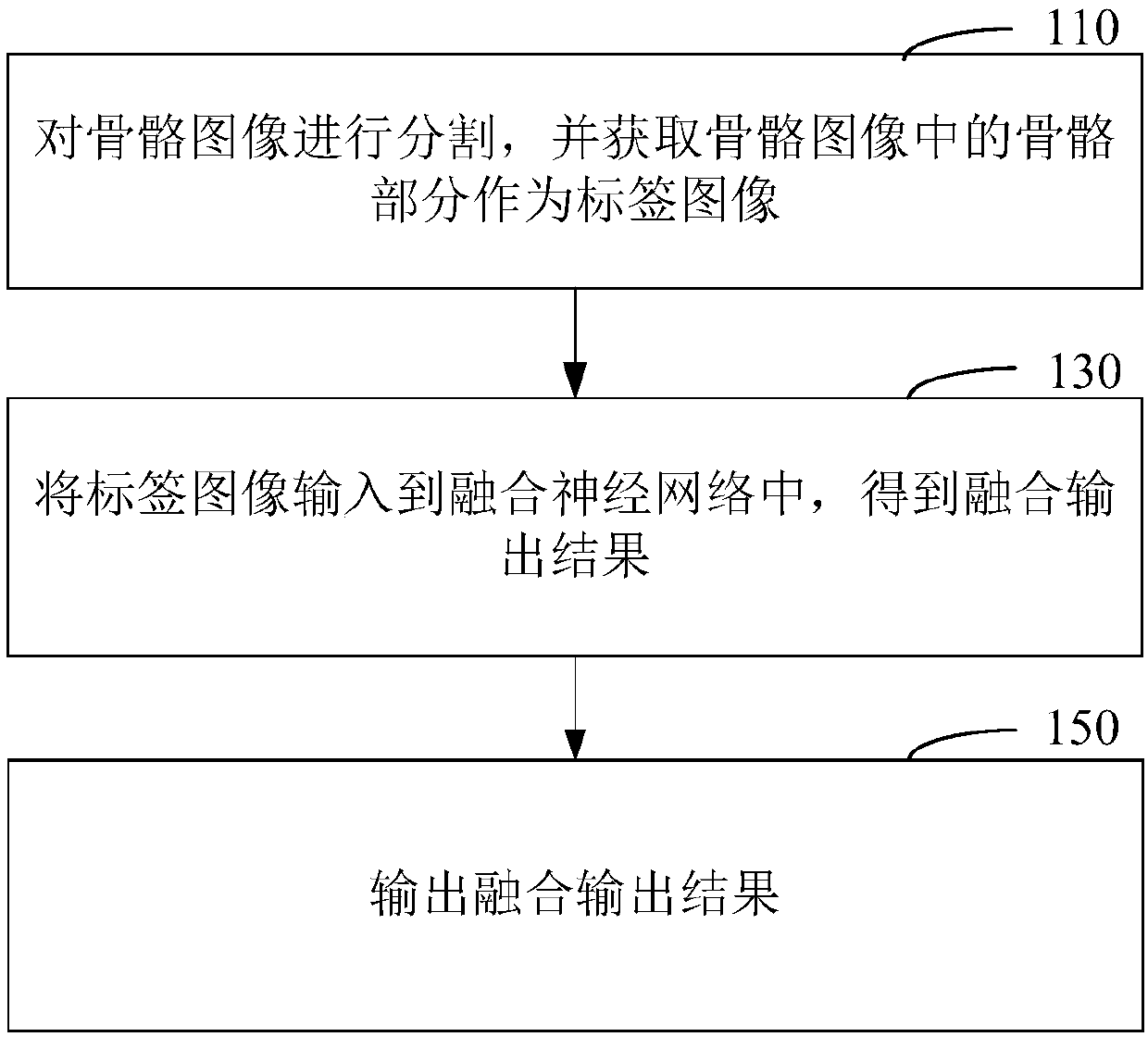
[0027] figure 1 It is a flow chart of a skeleton image processing method shown according to an exemplary embodiment. Such as figure 1 As shown, the bone image processing method may include the following steps.

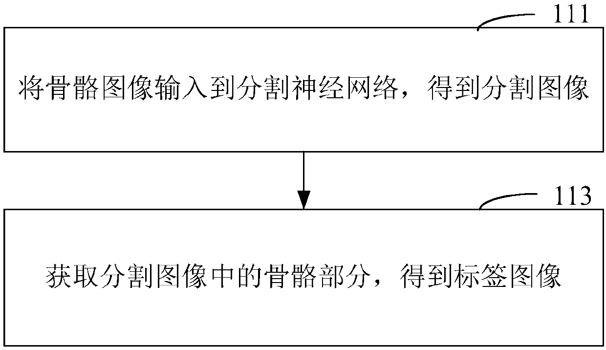
[0028] In step 110, the skeleton image is segmented, and the skeleton part in the skeleton image is acquired as a label image.

[0029] Among them, the bone image is first obtained, whic...


PUM


Abstract

The present invention discloses a bone image processing method, an electronic device and a computer readable storage medium. The method includes the following steps: splitting a bone image, and acquiring a bone part in the bone image as a label image; inputting the label image into a fused neural network to obtain a fused output result, wherein the fused neural network includes a classification neural network and a detection neural network; and outputting the fused output result, wherein the fused output result includes the fracture location and fracture probability of the bone image. A doctorcan know the possible location of the fracture in the bone image according to the fused output result including the fracture type, location and probability of the bone image without full-coverage untargeted examination of the bone image, so the manual workload of the fracture detection is greatly reduced, and the rate of missed diagnosis is also greatly reduced.

Description

technical field [0001] The invention relates to the technical field of computerized tomography image processing, in particular to a bone image processing method, electronic equipment and a computer-readable storage medium. Background technique [0002] Nowadays, in addition to emergency surgery, trauma surgery, spinal surgery and other surgical departments, fracture detection sources also include pain diagnosis and treatment departments, tumor radiotherapy departments, and digestive tract departments. There are nearly a thousand sheets, which need to be checked one by one, and the workload of fracture detection is huge. [0003] In actual work, clinicians often have no time to record the patient's injury details or medical history description due to their busy work, so radiologists need to conduct non-targeted comprehensive examinations for patients, which is very unfavorable for the control of missed diagnosis rate. [0004] For clinicians, it is necessary to identify each...

Claims


Application Information

Patent Timeline
IPC IPC(8): G16H30/20G06T7/00G06T7/10
CPCG16H30/20G06T7/0012G06T7/10G06T2207/10081G06T2207/30008G06T2207/20081G06T2207/20084
Inventor 王立王建军杨姗田广野
Owner QINGDAO HISENSE MEDICAL EQUIP
Why Eureka
Try Eureka