
Ultrasonic image preprocessing system for diagnosing breast lumps and method for diagnosing breast lumps

An ultrasound image and preprocessing technology, applied in the field of image processing, can solve the problems such as the inability to display the detailed information or lesion information of the breast image, the blurring probability of the detailed information of the breast image processing result, and the differential processing of the breast image, etc. Clear information, reduced iterations, and improved anti-noise performance

Inactive Publication Date: 2019-11-19
赵旭东
View PDF3 Cites 4 Cited by
  • Summary
  • Abstract
  • Description
  • Claims
  • Application Information

AI Technical Summary

Problems solved by technology

[0003] At present, the processing of different types of breast images is limited to using the same set of breast image processing methods, and different breast images are not processed differently, which will lead to a greater probability of fuzzy details in the obtained breast image processing results
For example, when enhancing different types of breast images, only using a fixed set of breast image enhancement methods may lead to the problem that some breast images are under-enhanced, while other breast images are over-enhanced, and finally the detailed information of breast images or Lesion information cannot be displayed clearly

Method used


Image

  • Ultrasonic image preprocessing system for diagnosing breast lumps and method for diagnosing breast lumps
  • Ultrasonic image preprocessing system for diagnosing breast lumps and method for diagnosing breast lumps
  • Ultrasonic image preprocessing system for diagnosing breast lumps and method for diagnosing breast lumps

Examples


Embodiment Construction

[0058] In order to enable those skilled in the art to better understand the present invention, the technical solution of the present invention will be further described below in conjunction with the accompanying drawings and embodiments.

[0059] The extraction process is described in detail with reference to the accompanying drawings and practical examples. The following is a step-by-step introduction:

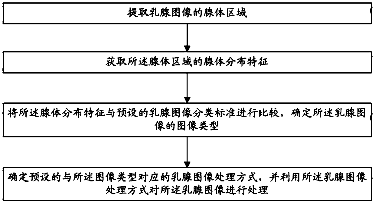
[0060] refer to figure 1 , figure 1 It is a flow chart of a breast image processing method provided in Embodiment 1 of the present invention, and the method includes:

[0061] S101: Extract a gland region of a breast image.

[0062]Since the glandular regions of the mammary gland image can reflect the distribution of the mammary glands, in this embodiment, the glandular regions of the mammary gland image first need to be extracted before processing the mammary gland image. There are many specific methods for extracting glandular regions of breast images. In this embodimen...


PUM


Abstract

The invention discloses an ultrasonic image preprocessing system and method for diagnosing breast lumps, and the method comprises the following steps: S1, extracting a gland area of a breast image, and S2, obtaining gland distribution features of the gland region; S3, determining the mammary gland density of a mammary gland image according to the proportion of the gland area in the foreground partof the mammary gland image; grading the breast density; S4, determining the image type of the mammary gland image in combination with the mammary gland density and the gland distribution characteristics; S5, determining a preset mammary gland image processing mode corresponding to the image type, and processing the mammary gland image by using the mammary gland image processing mode; S6, extracting a gland region of the mammary gland image; S7, determining the subareas of which the ratios are greater than a preset value as gland boundary areas, and calculating a segmentation threshold of eachgland boundary area; and S8, taking the average value of segmentation thresholds of gland boundary areas as a segmentation gland area threshold of the mammary gland image, and segmenting the gland boundary areas of the mammary gland image to obtain gland areas.

Description

technical field [0001] The invention belongs to the technical field of image processing, in particular to an ultrasonic image preprocessing system for diagnosing breast masses and a method for processing ultrasonic images. Background technique [0002] Since the composition of glands in different patients is different, and the distribution of breast glands is more varied, there may be large differences in mammogram images (hereinafter referred to as breast images) of different patients, and these differences may lead to discrepancies in breast images. Features such as edge information, contrast, and noise all vary. [0003] At present, the processing of different types of breast images is limited to using the same set of breast image processing methods, and different breast images are not processed differently, which will lead to a greater probability of fuzzy details in the obtained breast image processing results. For example, when enhancing different types of breast imag...

Claims


Application Information

Patent Timeline
Patent Type & Authority Applications(China)
IPC IPC(8): G06T7/00G06T7/11
CPCG06T7/0012G06T2207/10116G06T2207/10132G06T2207/20081G06T2207/30068G06T7/11
Inventor 赵旭东
Owner 赵旭东
Learn More