
Attenuation map for combined magnetic resonance-positron emission tomography

A technology of positron emission and tomography, applied in computerized tomography scanners, instruments for radiological diagnosis, applications, etc., can solve the problems of prolonging the total measurement duration and not being individualized, so as to achieve short measurement duration and prevent The effect of disappearing signal and improving comfort

Active Publication Date: 2021-01-19
SIEMENS HEALTHCARE GMBH
View PDF5 Cites 0 Cited by
  • Summary
  • Abstract
  • Description
  • Claims
  • Application Information

AI Technical Summary

Problems solved by technology

Regarding two imaging measurements in MRT, may require reciprocating movement of the table on which the patient lies, thus prolonging the total measurement duration
[0008] Furthermore, the sequence parameters of the two imaging measurements in MRT are often not individually adapted to the patient, in particular to the patient's posture and / or anatomy

Method used


Image

  • Attenuation map for combined magnetic resonance-positron emission tomography
  • Attenuation map for combined magnetic resonance-positron emission tomography
  • Attenuation map for combined magnetic resonance-positron emission tomography

Examples


Embodiment Construction

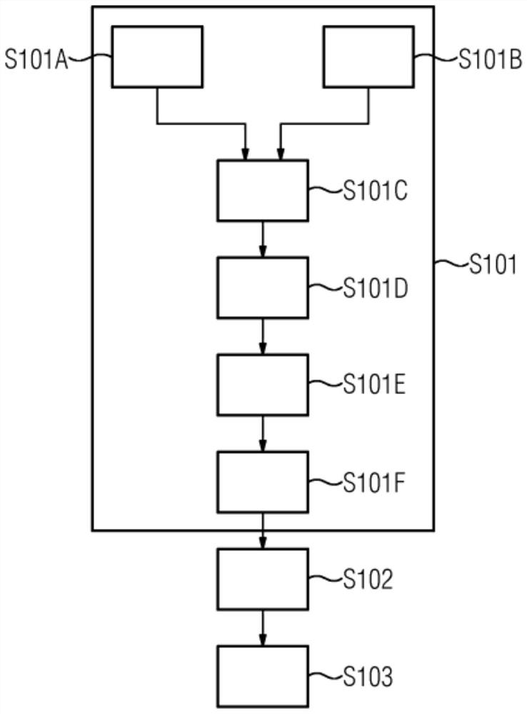
[0053] figure 1 A flowchart showing a method in a first embodiment for providing an attenuation map of a patient P suitable for correcting the patient's in- PET data detected in combined magnetic resonance-positron emission tomography (MRT-PET imaging).

[0054] Method step S101 represents the detection of magnetic resonance data in at least one imaging measurement in a magnetic resonance-positron emission tomography facility 10 .

[0055] Method step S101A represents the detection of first projection data of the patient P along its substantially axial direction.

[0056] Method step S101B represents detecting second projection data of the patient P along a substantially axial direction of the patient P.

[0057] Method step S101C represents obtaining third projection data by means of combining the first projection data of the patient P and the second projection data of the patient P.

[0058] Method step S101D represents deriving a quantitative map of the patient P using t...


PUM


Abstract

The present invention relates to a method for providing an attenuation map of a patient, a method for providing a corrected PET detected in a combined magnetic resonance-positron emission tomography scan of a patient in a magnetic resonance-positron emission tomography facility. data, a computerized positron emission tomography facility and an associated computer program product. The method according to the invention for providing an attenuation map of a patient is suitable for correcting PET data of a patient detected in a combined magnetic resonance-positron emission tomography scan in a magnetic resonance-positron emission tomography facility, said method comprising The following steps: detecting magnetic resonance data in a magnetic resonance-positron emission tomography facility, using the magnetic resonance data to derive an attenuation map of the patient, and providing an attenuation map of the patient for use in correcting the patient's combined magnetic resonance-positron emission tomography scan Attenuation plot of PET data detected in .

Description

technical field [0001] The present invention relates to a method for providing an attenuation map of a patient A method for providing corrected PET data detected in a combined magnetic resonance-positron emission tomography scan of a patient in a magnetic resonance-positron emission tomography facility, a magnetic resonance-positron emission tomography Tomography facility and an associated computer program product. Background technique [0002] Attenuation maps are typically created in combined magnetic resonance-positron emission tomography (MRT-PET imaging) at a magnetic resonance-positron emission tomography facility (MRT-PET facility). MRT-PET typically has a magnetic resonance tomography (MRT) and a positron emission tomography (PET). MRT-PET imaging typically includes at least one imaging measurement in MRT (MRT imaging) and at least one imaging measurement in PET (PET imaging). [0003] The attenuation map can be derived on the basis of magnetic resonance data (MR...

Claims


Application Information

Patent Timeline
Patent Type & Authority Patents(China)
IPC IPC(8): G16H30/40A61B5/055A61B6/03
CPCG16H30/40A61B5/055A61B5/0035A61B6/037A61B6/5247A61B6/582G01R33/481G16H40/63G16H30/20A61B6/4417A61B6/5258A61B5/7257G01T1/1603G01R33/56563G01R33/56572
Inventor 多米尼克·保罗马里奥·策勒弗拉维奥·卡林奇
Owner SIEMENS HEALTHCARE GMBH
Learn More