
Endoscope diagnosis support system, storage medium and endoscope diagnosis support method

A diagnosis aid and endoscope technology, applied in the direction of endoscope, diagnosis, TV system components, etc., can solve problems that prevent users from paying attention to medical images

Active Publication Date: 2022-05-17
OLYMPUS CORP
View PDF28 Cites 0 Cited by
  • Summary
  • Abstract
  • Description
  • Claims
  • Application Information

AI Technical Summary

Problems solved by technology

[0003] However, in the conventional image processing apparatus, before the user notices the lesion appearing in the medical image, a mark is displayed on the part of the suspected lesion, which sometimes prevents the user from paying attention to the medical image.

Method used


Image

  • Endoscope diagnosis support system, storage medium and endoscope diagnosis support method
  • Endoscope diagnosis support system, storage medium and endoscope diagnosis support method
  • Endoscope diagnosis support system, storage medium and endoscope diagnosis support method

Examples


no. 1 Embodiment approach

[0025] (structure)

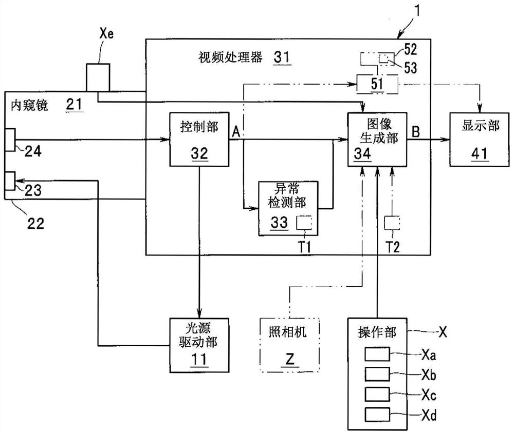
[0026] figure 1 It is a block diagram showing a configuration example of the endoscope diagnosis support system 1 according to the first embodiment of the present invention. exist figure 1 In , the illustration of the signal line connecting the operation part X and the control part 32 for setting the observation mode is omitted.

[0027] The endoscope diagnosis support system 1 has a light source drive unit 11 , an endoscope 21 , a video processor 31 , a display unit 41 and an operation unit X. As shown in FIG. The light source driving unit 11 is connected to the endoscope 21 and the video processor 31 . The endoscope 21 and the operation unit X are connected to a video processor 31 . The video processor 31 is connected to a display unit 41 .

[0028] The light source driving unit 11 is a circuit that drives the lighting unit 23 provided at the distal end of the insertion unit 22 of the endoscope 21 . The light source driving unit 11 is connected to ...

no. 2 Embodiment approach

[0109] In the first embodiment and Modifications 1 to 5 of the first embodiment, the subregion B2 displayed the detection position of the abnormality candidate region L, but an enlarged image E of the abnormality candidate region L may be displayed.

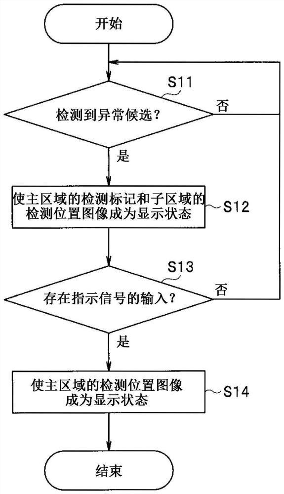
[0110] Figure 13 It is a flowchart showing an example of display image generation processing of the endoscope diagnosis support system 1 according to the second embodiment of the present invention. Figure 14 It is a diagram showing a configuration example of a display image B on the display unit 41 of the endoscope diagnosis support system 1 according to the second embodiment of the present invention. In this embodiment, descriptions of the same configurations as those of other embodiments and modifications are omitted.

[0111] The operation of the endoscope diagnosis support system 1 of the second embodiment will be described.

[0112] S31 to S33 are the same as S11 to S13, so the description thereof will be omitted.

[01...


PUM


Abstract

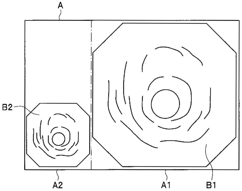
The endoscopic diagnosis support system (1) includes: an abnormality detecting unit (33) for detecting an abnormality candidate region (L) based on an endoscopic image (A) obtained by imaging the inside of a subject with an imaging unit (24) , and output the detection result; and an image generating section (34) that generates a display image (B) that is divided into a main area (B1) and a sub-area smaller than the main area (B1) ( B2), the endoscopic image (A1) is arranged in the main area (B1), and the abnormality detection image (Ma) indicating that the abnormality candidate area (L) is detected is arranged in the peripheral part of the main area (B1) according to the detection result .

Description

technical field [0001] The present invention relates to an endoscope diagnosis assistance system, an endoscope diagnosis assistance program and an endoscope diagnosis assistance method. Background technique [0002] Conventionally, there is a technique for performing image processing on medical images, marking and displaying portions that meet predetermined conditions. For example, Japanese Patent Application Laid-Open No. 2004-159739 discloses an image processing device that performs medical imaging from an X-ray CT device, a magnetic resonance imaging device, an ultrasonic diagnostic device, an X-ray imaging device, etc., in order to assist in diagnosis. Image processing of images, labeling and displaying parts of suspected lesions. [0003] However, in the conventional image processing apparatus, before the user notices the lesion appearing in the medical image, a mark is displayed on a portion suspected of being a lesion, which may prevent the user from paying attention...

Claims


Application Information

Patent Timeline
Patent Type & Authority Patents(China)
IPC IPC(8): A61B1/045
CPCA61B1/045A61B1/00045A61B1/0676A61B1/05A61B1/0005G06T7/0012G06T2207/10068G06T2207/30096G06T2207/20084G02B23/24A61B1/000094A61B1/000096A61B1/0655H04N23/555G06T2207/30204
Inventor 北村诚谷口胜义合渡大和河野隆志上山都士也神田大和
Owner OLYMPUS CORP
Learn More