Method and device for liver and liver tumor image segmentation

A liver tumor and image segmentation technology, which is applied in the field of medical image processing, can solve the problems of model fitting, large amount of parameters, and large amount of calculation, and achieve the effects of reducing false positives, accurate segmentation, and reducing the amount of model parameters

Pending Publication Date: 2020-05-19
BEIJING INSTITUTE OF TECHNOLOGYGY
View PDF4 Cites 10 Cited by
  • Summary
  • Abstract
  • Description
  • Claims
  • Application Information

AI Technical Summary

Problems solved by technology

Although 3DFCN makes full use of the spatial structure information of volume data, it has a large amount of parameters and a large amount of calculation. It is ...

Method used


Image

  • Method and device for liver and liver tumor image segmentation
  • Method and device for liver and liver tumor image segmentation
  • Method and device for liver and liver tumor image segmentation

Examples


Embodiment Construction

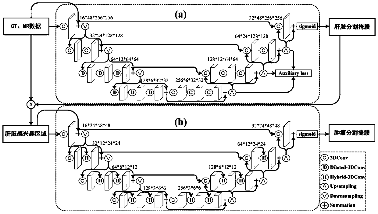
[0023] Such as Figure 4 As shown, the image segmentation method of the liver and liver tumors includes the following steps:

[0024] (1) Obtain abdominal magnetic resonance images;

[0025] (2) Use the liver model to determine the region of interest. The liver model is Dial3DResUNet (hole three-dimensional residual U-shaped neural network), which combines long and short-range skip connection structure and mixed hole convolution to fully capture the global structural information of the image for accurate liver segmentation;

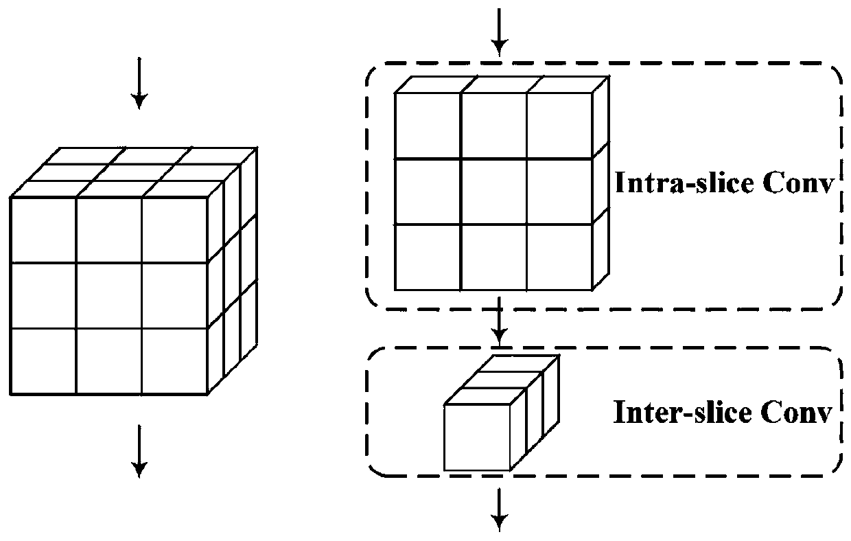
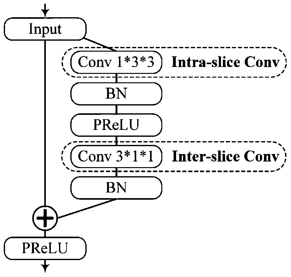
[0026] (3) Use the liver tumor model for fine segmentation to reduce false positives. The liver tumor model is H3DNet (Hybrid 3D Convolutional Neural Network), which consists of Hybrid-3D (Hybrid 3D) convolution, which effectively extracts 3D features of liver tumors. At the same time, the amount of model parameters is greatly reduced, and the difficulty of model optimization and the risk of overfitting are reduced.

[0027] The present invention uses ...


PUM


Abstract

The invention discloses a method and a device for liver and liver tumor image segmentation. The method and the device can effectively and accurately segment liver and liver tumors in different modes.The method comprises the following steps: (1) an abdomen magnetic resonance image is acquired; (2) a liver model is used to determine a region of interest, and the liver model is Dial3DResUNet, and iscombined with a long-short distance jump connection structure and mixed hole convolution to fully capture image global structure information so as to perform accurate liver segmentation; and (3) a liver tumor model is used for fine segmentation to reduce false positive results, wherein the liver tumor model is H3DNet and is composed of Hybrid 3D convolution. The model parameter quantity is greatly reduced while the liver tumor three-dimensional features are effectively extracted, and the model optimization difficulty and the overfitting risk are reduced.

Description

technical field [0001] The present invention relates to the technical field of medical image processing, in particular to a liver and liver tumor image segmentation method, and also relates to a liver and liver tumor image segmentation device. Background technique [0002] Liver cancer is the sixth most prevalent and second most lethal cancer in the world. It was responsible for 782,000 deaths worldwide in 2012 and 810,500 in 2015. Liver tumor segmentation is an important step in the preoperative diagnosis of liver cancer, the formulation of surgical plans, and the evaluation of postoperative efficacy. However, manually segmenting the liver and liver tumors is time-consuming and labor-intensive, and requires doctors to accumulate a lot of experience. Therefore, automatic liver and liver tumor segmentation is very necessary to assist doctors in their daily work. [0003] However, the automatic segmentation of liver and tumor is very challenging. The contrast between the l...

Claims


Application Information

Patent Timeline
IPC IPC(8): G06T7/00G06T7/11
CPCG06T7/0012G06T7/11G06T2207/10081G06T2207/10088G06T2207/20081G06T2207/20084G06T2207/30056
Inventor 杨健宋红范敬凡张超逸王涌天
Owner BEIJING INSTITUTE OF TECHNOLOGYGY
Why Eureka
Try Eureka