Environment monitoring method and device for vehicle-mounted computed tomography equipment
A technology of on-board computer and tomography, which is applied in the direction of measuring devices, measurement value indicators, instruments, etc.
- Summary
- Abstract
- Description
- Claims
- Application Information
AI Technical Summary
Problems solved by technology
Method used
Image
Examples
Embodiment 1
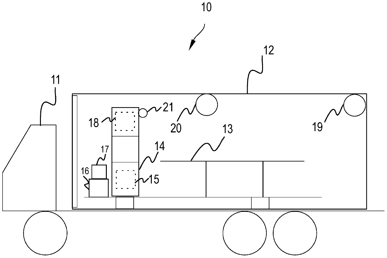
[0086] The processor in the control host 16 via the temperature sensor ( figure 2 not shown in ) to obtain the temperature of the X-ray tube 18 (for example, the anode temperature).
[0087] In an optional embodiment, when the processor finds that the temperature of the X-ray tube 18 is too high (such as exceeding the upper limit of the temperature of the X-ray tube), the processor can send a message containing X-rays to a remote server via the wireless interface 17. An abnormal alarm message of the temperature value of the tube 18, the abnormal alarm message further includes a field for identifying the faulty object (that is, the X-ray tube 18). After receiving the abnormal alarm message, the server determines that the temperature of the X-ray tube 18 is too high based on this field, and sends an X-ray tube cooling instruction to the wireless interface 17 . The processor in the control host 16 receives the X-ray tube cooling instruction via the wireless interface 17, and co...
Embodiment 2
[0090] The processor in the control host 16 acquires the ambient temperature value in the compartment 12 via the temperature sensor 19 arranged in the compartment 12 . In an optional embodiment, when the processor finds that the ambient temperature value in the compartment 12 is too high (such as exceeding the upper limit of the compartment temperature), the processor sends the temperature information containing the compartment 12 to a remote server via the wireless interface 17. Value abnormal alarm message, the fault alarm message further includes a field for identifying the fault object (that is, the compartment 12). After receiving the abnormal alarm message, the server determines that the temperature of the compartment 12 is too high based on this field, and then sends a compartment cooling instruction to the wireless interface 17 . The processor in the control host 16 receives the compartment cooling command via the wireless interface 17, and cools the compartment 12 based...
Embodiment 3
[0093] The processor in the control host 16 acquires the temperature of the X-ray tube 18 (for example, the anode temperature) via the temperature sensor arranged in the X-ray tube 18 of the scanning frame 14, and obtains the temperature of the compartment 12 via the temperature sensor 19 arranged in the compartment 12. The ambient temperature value in . When the processor finds that the temperature of the X-ray tube 18 is too high (such as exceeding the upper limit of the temperature of the X-ray tube) and the ambient temperature value in the compartment 12 is too high (such as exceeding the upper limit of the temperature of the compartment), it is determined that the X-ray tube 18 and the compartment The environment of 12 is abnormal. Moreover, the processor determines that the abnormal state of the temperature of the X-ray tube is the same as the abnormal state of the temperature of the compartment 12, both of which are too high. The control host generates an instruction f...
PUM
 Abstract
Description
Claims
Application Information
 - R&D
- Intellectual Property
- Life Sciences
- Materials
- Tech Scout
- Unparalleled Data Quality
- Higher Quality Content
- 60% Fewer Hallucinations
Browse by: Latest US Patents, China's latest patents, Technical Efficacy Thesaurus, Application Domain, Technology Topic, Popular Technical Reports.
© 2025 PatSnap. All rights reserved.Legal|Privacy policy|Modern Slavery Act Transparency Statement|Sitemap|About US| Contact US: help@patsnap.com


