Veterinary fixing device for imaging diagnosis of feline

A feline, fixed device technology, applied in the directions of diagnosis, medical science, patient positioning for diagnosis, etc., can solve problems such as difficult positioning, unable to display images, etc., to achieve the effect of light weight, reliability and safety

Inactive Publication Date: 2020-07-31
杭州宸熙辉创网络科技有限公司
View PDF0 Cites 0 Cited by
  • Summary
  • Abstract
  • Description
  • Claims
  • Application Information

AI Technical Summary

Problems solved by technology

However, given the specific anatomical shape of felines, it was always difficult to position them correctly inside the machine for CT scanning
Specifically, the anatomical shape of the feline makes it difficult to position the feline properly in the prone or supine position becaus...

Method used


Image

  • Veterinary fixing device for imaging diagnosis of feline
  • Veterinary fixing device for imaging diagnosis of feline
  • Veterinary fixing device for imaging diagnosis of feline

Examples


Embodiment Construction

[0020] Embodiments of the invention are described in detail below, examples of which are illustrated in the accompanying drawings. The embodiments described below by referring to the figures are exemplary and are intended to explain the present invention and should not be construed as limiting the present invention.

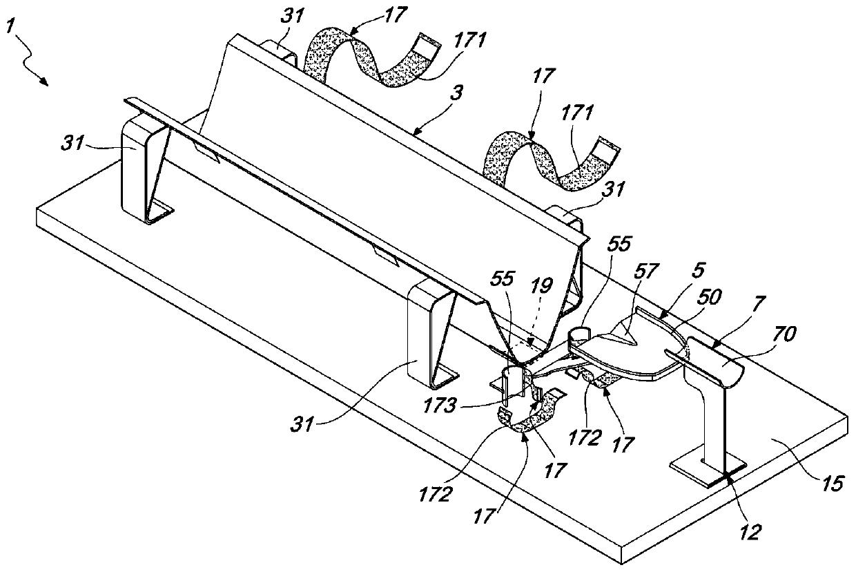
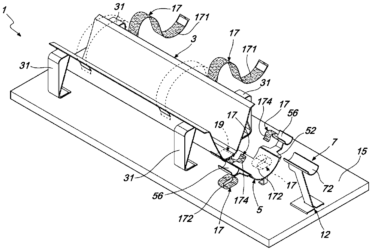
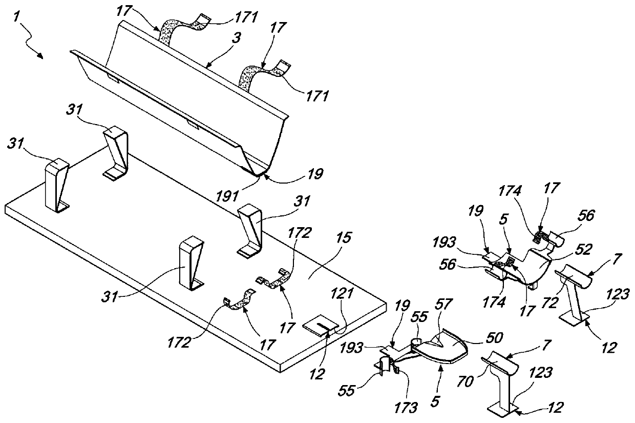
[0021] Such as Figure 1-5 As shown, a veterinary feline imaging diagnostic fixation device 1 of the present invention includes a trunk support frame 3 for accommodating a torso 101 of a feline 100 and a skull support frame 5 for supporting a head 103 of a feline 100 .

[0022] The immobilization device 1 has a first configuration position in which the feline 100 is supported in a prone position P and a second configuration position in which the feline 100 is supported in a supine position S, figure 1 with Figure 4 An immobilization device 1 suitable for supporting a cat 102 in a prone position P is shown, figure 2 with Figure 5 An immobilization device 1 ...


PUM


Abstract

The invention belongs to the veterinary field and discloses a veterinary fixing device for imaging diagnosis of a feline. The fixing device comprises a trunk support frame for accommodating the trunkof the feline, a skull support frame for supporting the head of the feline, a support element, multiple bands and a base, wherein the skull support frame comprises a first skull support frame and a second skull support frame, and the support element comprises a first support element and a second support element for fixation in a prostrate position P and a supine position S of the feline. The fixing device correctly positions the feline through the axis of a CT machine, acquires display of a diagnosis image with symmetric left and right sides of the feline, does not cause artifacts in the diagnosis image, is light in weight, durable and disinfectable, can support different sizes of felines and ensures the reliability and safety in use to the maximum extent.

Description

technical field [0001] The invention relates to the field of veterinary medicine, in particular to a veterinary feline imaging diagnosis fixation device. Background technique [0002] Currently, there is an increasing need for imaging diagnostic examinations in felines, especially computed axial tomography, in order to schedule appropriate interventions, such as veterinary surgery. For example, common pet domestic cats are placed directly on the CT scanning workbench during imaging diagnosis. However, given the specific anatomical shape of felines, it has always been difficult to position them correctly inside the machine for CT scanning. Specifically, the anatomical shape of the feline makes it difficult to position the feline properly in the prone or supine position because the anatomical shape of the feline torso has a generally oval cross-section, forcing the operator to position the feline in the The edges of the table, ie the torso and head are rotated relative to th...

Claims


Application Information

Patent Timeline
IPC IPC(8): A61B6/04A61B6/03
CPCA61B6/032A61B6/04A61B2503/40
Inventor 徐立勇
Owner 杭州宸熙辉创网络科技有限公司
Why Eureka
Try Eureka