
Medical image enhancement method and computer readable storage medium

An image and grayscale image technology, applied in the field of medical image enhancement methods and computer-readable storage media, can solve the problem of low detection rate of lesions

Pending Publication Date: 2020-08-04
SHANGHAI UNITED IMAGING INTELLIGENT MEDICAL TECH CO LTD
View PDF10 Cites 3 Cited by
  • Summary
  • Abstract
  • Description
  • Claims
  • Application Information

AI Technical Summary

Problems solved by technology

[0005] Based on this, it is necessary to provide a medical image enhancement method and a computer-readable storage medium for the problem that the detection rate of special types of breast lesions in traditional technologies is still low

Method used


Image

  • Medical image enhancement method and computer readable storage medium
  • Medical image enhancement method and computer readable storage medium
  • Medical image enhancement method and computer readable storage medium

Examples


Embodiment Construction

[0074] In order to make the purpose, technical solutions, and advantages of this application clearer, the following further describes this application in detail with reference to the accompanying drawings and embodiments. It should be understood that the specific embodiments described here are only used to explain the application, and not used to limit the application.

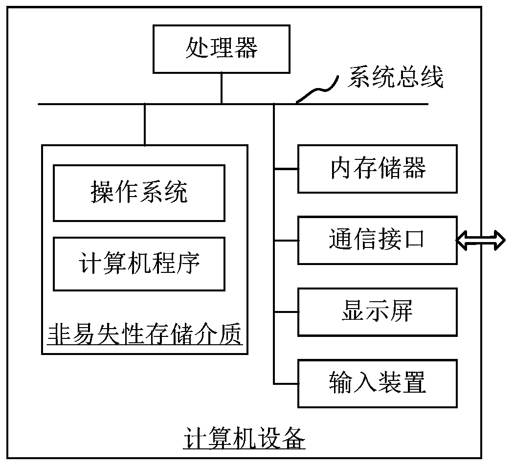
[0075] The medical image enhancement method provided by the embodiments of this application can be applied to figure 1 Computer equipment shown. The computer device includes a processor and a memory connected through a system bus, and a computer program is stored in the memory. When the processor executes the computer program, the steps of the following method embodiments can be executed. Optionally, the computer equipment may also include a communication interface, a display screen, and an input device. Among them, the processor of the computer device is used to provide calculation and control capabilities. Th...


PUM


Abstract

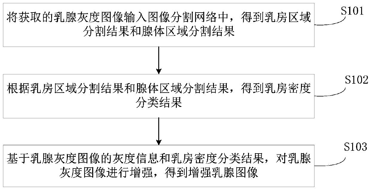
The invention relates to a medical image enhancement method and a computer readable storage medium. The method comprises the following steps: inputting an acquired mammary gland grayscale image into an image segmentation network to obtain a breast region segmentation result and a gland region segmentation result; calculating the area proportion of the gland region in the breast region according tothe breast region segmentation result and the gland region segmentation result; obtaining a breast density classification result through the area ratio; and enhancing the mammary gland grayscale image based on the grayscale information of the mammary gland grayscale image and the breast density classification result to obtain an enhanced mammary gland image. According to the method, mammary glandgrayscale images are classified; different enhancement strategies are applied to images with different breast densities, so that focus areas in enhanced mammary gland images obtained from different mammary gland grayscale images can be more obvious, the focus areas can be detected more easily in the focus detection process, and the focus detection rate of different types of mammary gland grayscale images is increased.

Description

Technical field [0001] This application relates to the field of image processing technology, and in particular to a medical image enhancement method and a computer-readable storage medium. Background technique [0002] Breast cancer is one of the malignant tumors that seriously endanger women’s health, and it is also the main cause of death from cancer in women. At present, there is no effective means to prevent breast cancer in medicine. Therefore, increasing the detection rate of breast cancer imaging examinations to treat symptomatically, Reducing the mortality rate of breast cancer is of great significance. [0003] Currently, mammography is the mainstream breast cancer examination method, but its diagnostic performance is affected by breast density imaging, especially the detection rate of dense breast gland lesions has limitations. Therefore, it is necessary to enhance mammograms to improve the detection rate of lesions. In traditional technology, the same image enhancement...

Claims


Application Information

Patent Timeline
Patent Type & Authority Applications(China)
IPC IPC(8): G06T5/00G06T7/62G06T7/90G06T7/11G06T7/00G06K9/62
CPCG06T7/62G06T7/90G06T7/11G06T7/0012G06T2207/10116G06T2207/30068G06F18/2414G06T5/73
Inventor 李哲人郑介志
Owner SHANGHAI UNITED IMAGING INTELLIGENT MEDICAL TECH CO LTD
Learn More