
Artificial intelligence multimode imaging analysis system

An artificial intelligence and multi-mode imaging technology, applied in echo tomography, medical science, patient positioning for diagnosis, etc., can solve the problems of patients lying on the examination bed and patients getting up, and achieve the effect of accelerated diagnosis

Inactive Publication Date: 2020-10-16
丁佳丽
View PDF7 Cites 0 Cited by
  • Summary
  • Abstract
  • Description
  • Claims
  • Application Information

AI Technical Summary

Problems solved by technology

[0004] However, during the detection process, there are often some weak patients or elderly people, and the height of the examination bed is generally higher than the normal bed, so it is difficult for the patient to lie on the examination bed, and it is difficult for the patient to get up after the examination. It is very difficult, so we propose an artificial intelligence multimodal imaging analysis system

Method used


Image

  • Artificial intelligence multimode imaging analysis system
  • Artificial intelligence multimode imaging analysis system
  • Artificial intelligence multimode imaging analysis system

Examples


Embodiment 1

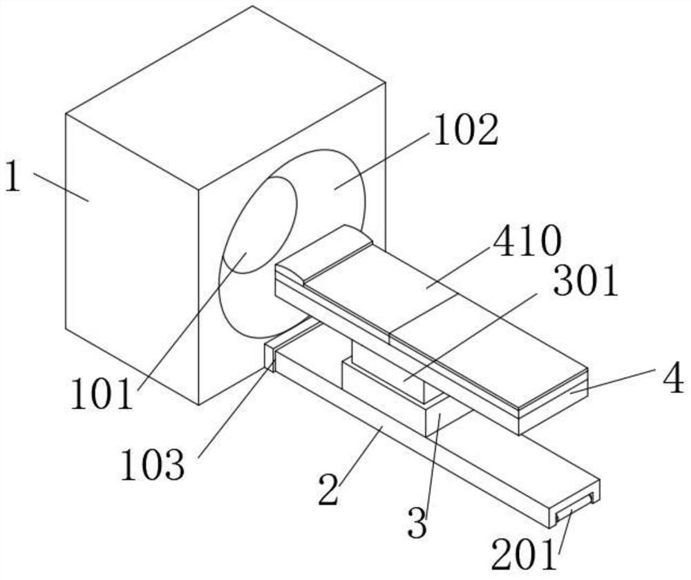
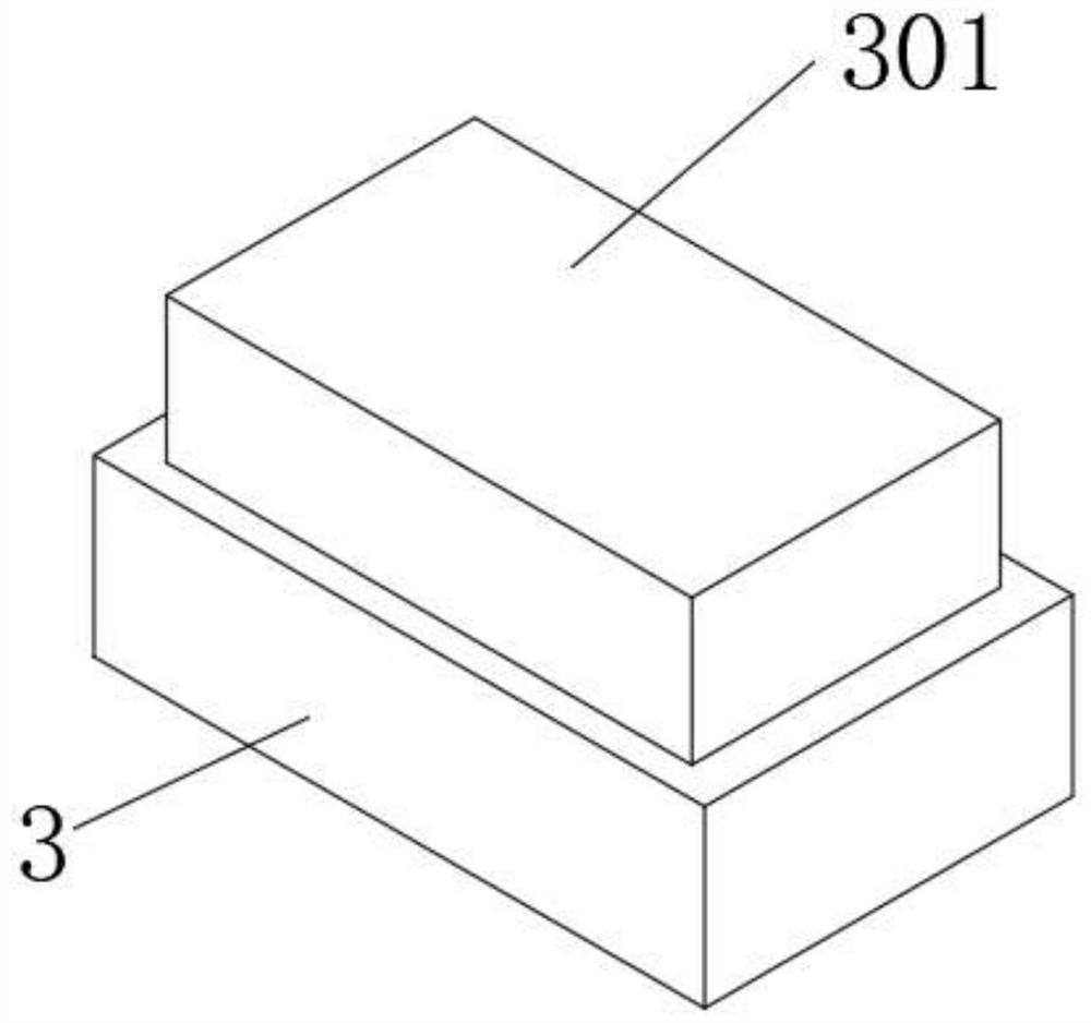
[0037] Embodiment 1: The embodiment of the present invention discloses an artificial intelligence multimodal imaging analysis system, such as Figure 1-7 As shown, the imager 1 is included, and the inside of the imager 1 is provided with a detection chamber 101. The inside of the detection chamber 101 is provided with an in vitro detector, and the front of the imager 1 is provided with an inlet 102, and the detection chamber 101 and the inlet 102 are connected. There is a power bin 103 at the bottom of the front of the imager 1, and the inside of the power bin 103 is provided with a driving mechanism and is connected to the base 2 through the driving mechanism transmission. The driving mechanism is a structure in an existing device, and the present invention will not repeat them;

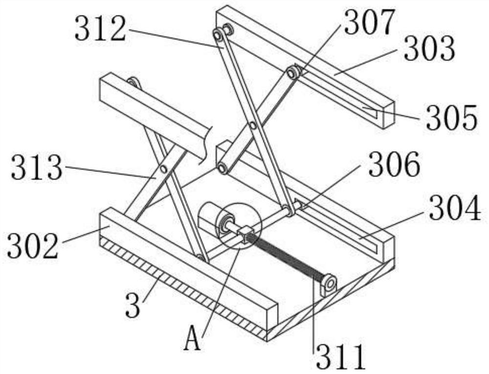
[0038] The upper surface of the base 2 is fixedly connected with a first protective frame 3, and the inner wall of the first protective frame 3 is slidably connected with a second protective frame 30...

Embodiment 2

[0049] Embodiment 2: The embodiment of the present invention discloses an artificial intelligence multimodal imaging analysis system, such as figure 1 and Figure 8 As shown, the following analysis steps are included:

[0050] S1 Positron tomography: First, the patient injects a small amount of positron nuclide tracer, and then the patient lies on the fixed plate 401 and the movable plate 402, and the driving mechanism inside the power chamber 103 drives the base 2 to move toward the inside of the power chamber 103 , until the patient enters the detection chamber 101, and the in vitro detector installed inside the detection chamber 101 detects the human body;

[0051] S2 multi-mode PET imaging: the in vitro detector detects the distribution of positron nuclides in various organs of the human body, and performs imaging by computerized tomography;

[0052] S3 artificial intelligence analysis: artificial intelligence quickly and effectively analyzes the distribution of positron...


PUM


Abstract

The invention discloses an artificial intelligence multimode imaging analysis system, and relates to the field of multimode imaging. The artificial intelligence multimode imaging analysis system comprises an imager, wherein a detection bin is formed in the imager; a power bin is arranged at the bottom of the front face of the imager; the power bin is in transmission connection with a base; a firstprotection frame is fixedly connected to the upper surface of the base, and a second protection frame is slidably connected to the inner wall of the first protection frame; a lifting mechanism is arranged between the first protection frame and the second protection frame; an examination bed is fixedly connected to the top of the second protection frame; a fixed plate is fixedly connected to the upper surface of the examination bed; a movable plate is rotationally connected to the fixed plate through a rotating shaft; a cavity is formed in the upper surface of the examination bed; and a transmission mechanism is arranged in the cavity. According to the artificial intelligence multi-mode imaging analysis system, the examination bed can be lifted, so that a weak patient can get on the bed conveniently, and meanwhile, the back of the patient can be slowly supported, so that the weak patient can get up conveniently.

Description

technical field [0001] The invention relates to the technical field of multi-mode imaging, in particular to an artificial intelligence multi-mode imaging analysis system. Background technique [0002] Multimodal imaging technology is to inject a small amount of positron nuclide tracers into the human body, and then use a special in vitro detector (PET) to detect the distribution of these positron nuclide in various organs of the human body. The method shows the physiological and metabolic functions of the main organs of the human body. [0003] Cell images contain more information than can be extracted by the human eye, and computer-assisted image analysis has the potential to make complex morphological information more accessible in daily diagnostic practice, improving prognosis and predicting patient stratification. [0004] However, during the detection process, there are often some weak patients or elderly people, and the height of the examination bed is generally highe...

Claims


Application Information

Patent Timeline
Patent Type & Authority Applications(China)
IPC IPC(8): A61B6/03A61B6/04
CPCA61B6/037A61B6/4057A61B6/0407
Inventor 丁佳丽王孟沈海波王文静
Owner 丁佳丽
Learn More