Liver cirrhosis ultrasonic image liver envelope extraction method based on digital image processing technology

A technology of ultrasonic images and digital images, applied in the field of medical imaging, can solve the problems of relying on experience and low efficiency

Active Publication Date: 2020-10-23
SHANGHAI UNIV OF ENG SCI
View PDF5 Cites 0 Cited by
  • Summary
  • Abstract
  • Description
  • Claims
  • Application Information

AI Technical Summary

Problems solved by technology

The traditional liver capsule extraction method mainly relies on manual marking by medical workers, which is ineffi

Method used


Image

  • Liver cirrhosis ultrasonic image liver envelope extraction method based on digital image processing technology
  • Liver cirrhosis ultrasonic image liver envelope extraction method based on digital image processing technology
  • Liver cirrhosis ultrasonic image liver envelope extraction method based on digital image processing technology

Examples


Embodiment Construction

[0046] The present invention will be described in detail below in conjunction with specific embodiments. The following examples will help those skilled in the art to further understand the present invention, but do not limit the present invention in any form. It should be noted that those skilled in the art can make several changes and improvements without departing from the concept of the present invention. These all belong to the protection scope of the present invention.

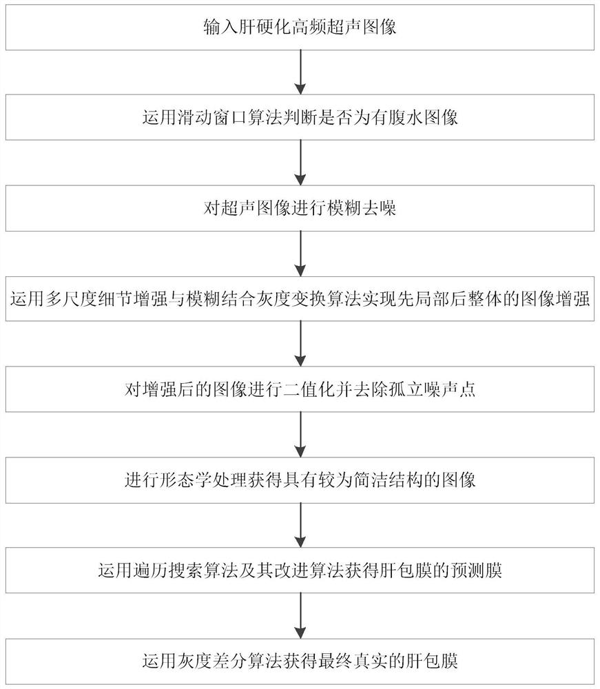
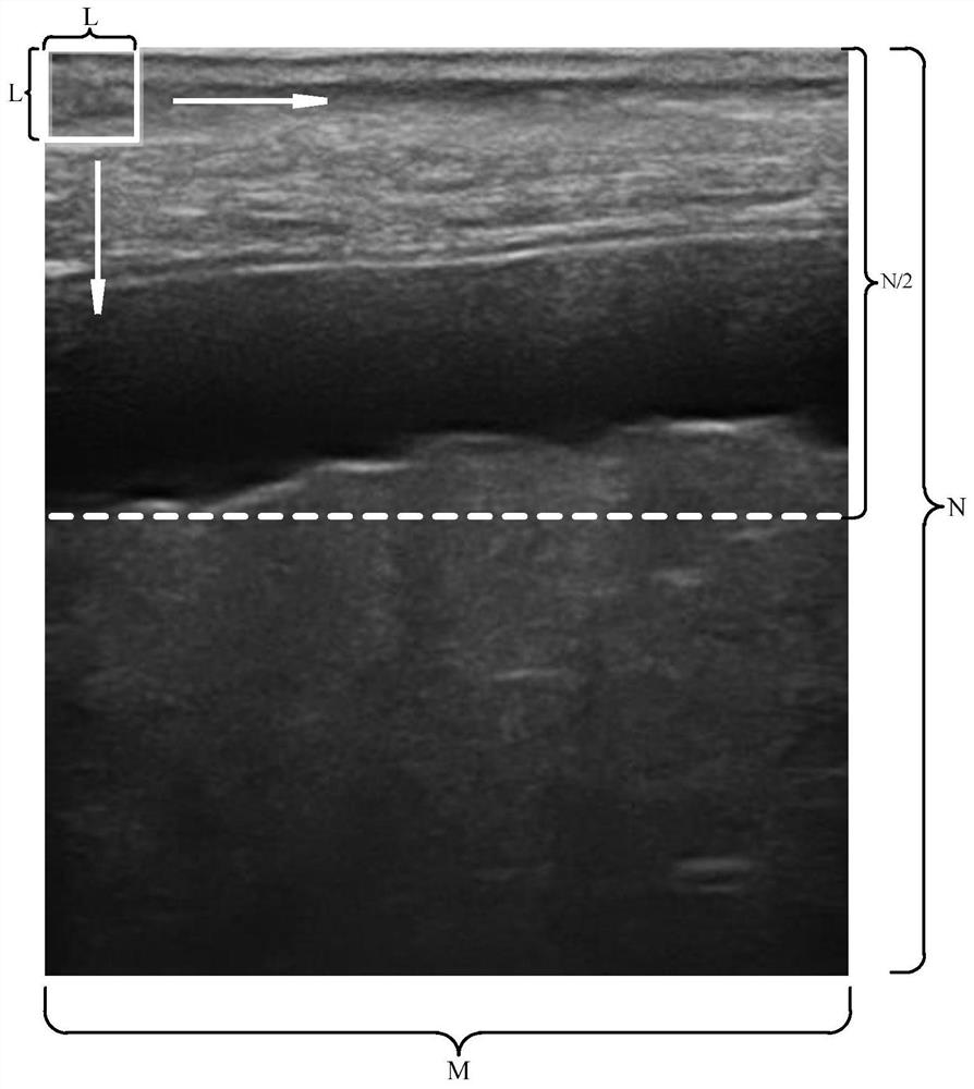
[0047] Embodiments of the present invention include a method for extracting the liver capsule from ultrasound images of liver cirrhosis based on digital image processing technology. The method is used to process superficial slice images of the liver. image. Liver superficial section images such as image 3 As shown, the image includes the liver capsule at the upper edge of the liver, the lower part of the image is the liver parenchyma, and the upper part is other organs.

[0048] Such as figure 1 as ...


PUM


Abstract

The invention provides a liver cirrhosis ultrasonic image liver envelope extraction method based on a digital image processing technology, which is used for processing a liver superficial section image, and comprises the following steps: traversing the upper part of an ultrasonic image by using a sliding window algorithm to identify whether a liver ascites area exists in the ultrasonic image or not; performing gaussian blur processing, binarization processing and morphological closed operation processing sequentially on the ultrasonic image to obtain a binary ultrasonic image; searching the processed binary ultrasonic image based on a traversal algorithm, and searching a pixel point set corresponding to the liver envelope to form a prediction film of the liver envelope; and fusing the prediction membrane into the original ultrasonic image, and removing pixel points of the pseudo membrane from the prediction membrane by adopting a gray difference algorithm to obtain a real membrane of the liver envelope. According to the invention, the prediction membrane and the real membrane of the liver envelope can be obtained at the same time, and a guarantee is provided for subsequent morphological characteristic analysis.

Description

technical field [0001] The invention relates to the field of medical imaging, in particular to a method for extracting a liver capsule from an ultrasound image of liver cirrhosis based on digital image processing technology. Background technique [0002] Liver cirrhosis, as a common chronic progressive disease, has serious harm to the human body. With the development of the disease, it can gradually lead to portal hypertension, end-stage liver disease, and even death. Therefore, for Early auxiliary diagnosis and treatment of liver cirrhosis is very important clinically. [0003] At present, the early diagnosis of liver cirrhosis using ultrasound images can be divided into two aspects, one is based on the analysis of the morphological characteristics of the liver capsule, and the other is based on the analysis of the texture characteristics of the liver parenchyma. Among them, how to quickly and accurately extract the liver envelope line is very important when analyzing the ...

Claims


Application Information

Patent Timeline
IPC IPC(8): G06T5/00G06T5/30G06T7/00G06T7/11G06T7/155G06T7/187G06T7/90
CPCG06T5/008G06T5/009G06T5/002G06T7/155G06T7/11G06T7/90G06T7/187G06T7/0012G06T5/30G06T2207/10132G06T2207/30056
Inventor 刘翔刘艺文王诗雨浦秀丽朱佳琦
Owner SHANGHAI UNIV OF ENG SCI
Why Eureka
Try Eureka