
Medical image segmentation method based on 3D dynamic edge insensitivity loss function

A loss function and medical image technology, applied in the field of medical image processing, can solve the problems affecting the generalization of the segmentation model and the high risk of model overfitting, so as to improve the generalization performance, improve the generalization and accuracy, and reduce the network The effect of the training parameters

Active Publication Date: 2020-11-20
FUDAN UNIV
View PDF3 Cites 2 Cited by
  • Summary
  • Abstract
  • Description
  • Claims
  • Application Information

AI Technical Summary

Problems solved by technology

When more attention is paid to the edge features of the region of interest in the limited training samples, although the indicators on the limited training samples can be continuously improved, the risk of over-fitting of the model is also getting higher and higher, which will inevitably affect the general performance of the segmentation model. Chemical

Method used


Image

  • Medical image segmentation method based on 3D dynamic edge insensitivity loss function
  • Medical image segmentation method based on 3D dynamic edge insensitivity loss function
  • Medical image segmentation method based on 3D dynamic edge insensitivity loss function

Examples


Embodiment 1

[0043] Embodiment 1 Using the method of the present invention to perform multi-region segmentation of the prostate

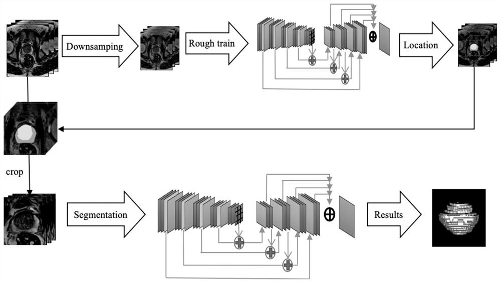
[0044] The medical image segmentation method based on the 3D dynamic edge insensitivity loss function provided by the present invention is end-to-end, and the specific implementation process of the embodiment is as follows:

[0045] Step 1. There are 68 cases in the training set data, which come from the ISBI data set. First, intensity normalization and histogram equalization are performed on the image, and the entire image is divided into many small blocks of pixels for nonlinear stretching, so that the local gray histogram is evenly distributed. In order for the network to correctly learn the spatial semantics, all MR voxels are resampled to a uniform size using third-order spline interpolation. Nearest neighbor interpolation is used for the corresponding segmentation annotations. Random transformations mainly including random rotation, shearing, scaling and...


PUM


Abstract

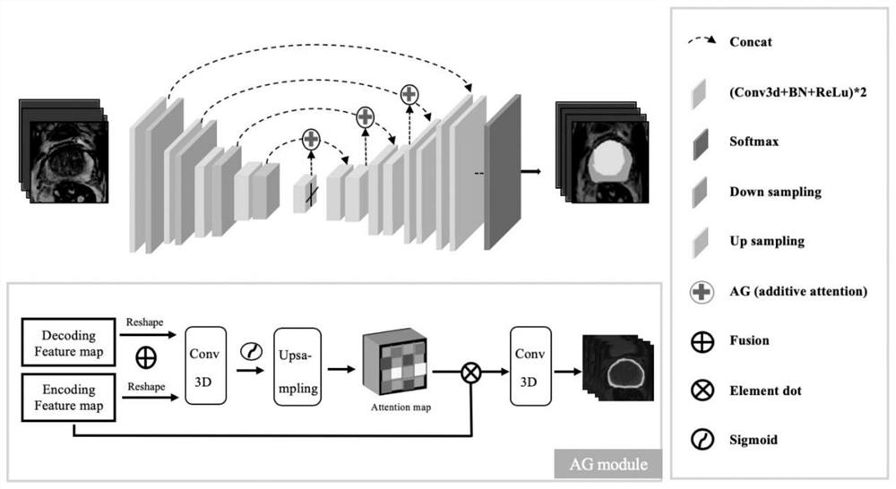
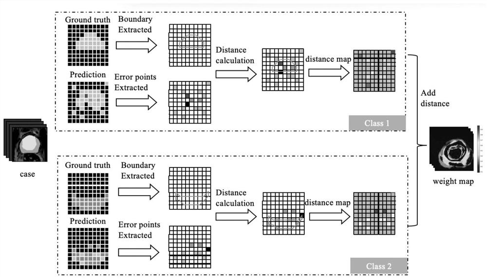
The invention belongs to the technical field of medical image processing, and particularly relates to a medical image segmentation method based on a 3D dynamic edge insensitivity loss function. The model adopts a dynamic edge insensitivity loss function, and the design principle of the loss function is as follows: in each iterative training process, the farther the prediction error pixel point isfrom the edge, the more sensitive the network is to the pixel point, and the larger the punishment weight is. In this way, the untrusted edge sensitivity can be reduced, and the influence of the edgeuncertainty of different expert annotation data on the model is reduced, and the generalization performance of the model for medical image segmentation is improved. And meanwhile, a U-net architecturebased on an attention mechanism is adopted, and the deviation of the network model is relatively small by fusing the weight distribution of the attention module self-adaptive feature map, so that theinfluence of annotation noise on model learning is reduced, and the generalization and accuracy of the medical image segmentation model are improved.

Description

technical field [0001] The invention belongs to the technical field of medical image processing, and in particular relates to a medical image segmentation method. Background technique [0002] The task of medical image segmentation is an important and essential work in clinical problems. With the rapid development of artificial intelligence, a large number of excellent algorithms have emerged, and the indicators of many challenge datasets have been continuously refreshed. It is currently recognized that the precise segmentation of edges is very important to improve the accuracy of segmentation, so there have been a lot of work to focus on the edge segmentation of medical images, and adding additional network branches to learn boundary features is one of the mainstream methods. There are also some works that add attention to edges in the loss function to achieve better segmentation results. [0003] The premise of these algorithms is that the ground-truth annotated edge segm...

Claims


Application Information

Patent Timeline
IPC IPC(8): G06T7/12G06T5/00G06T5/40
CPCG06T7/12G06T5/40G06T2207/20081G06T2207/20084G06T2207/20132G06T2207/30081G06T5/92
Inventor 章琛曦裘茗烟宋志坚
Owner FUDAN UNIV
Learn More