Automated hemostasis system for esophageal varices surgery

A technique for varicose veins and surgery, which is applied in the medical field and can solve problems such as injection hole bleeding

Active Publication Date: 2021-09-10
合肥中纳医学仪器有限公司
View PDF12 Cites 0 Cited by
  • Summary
  • Abstract
  • Description
  • Claims
  • Application Information

AI Technical Summary

Problems solved by technology

[0003] In order to solve the technical problem of injection hole bleeding in the existing varicose vein operation device, the present invention provides an automatic hemostasis system used in esophageal variceal operation

Method used


Image

  • Automated hemostasis system for esophageal varices surgery
  • Automated hemostasis system for esophageal varices surgery
  • Automated hemostasis system for esophageal varices surgery

Examples


Embodiment 1

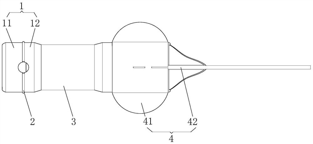
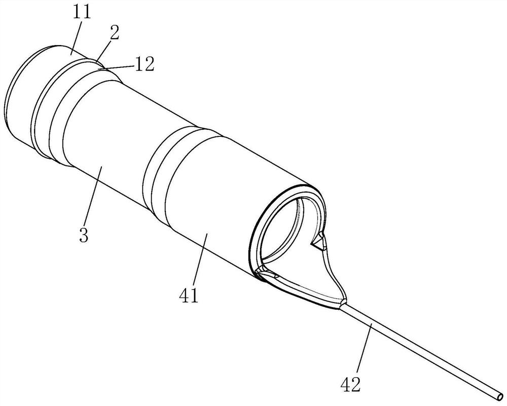
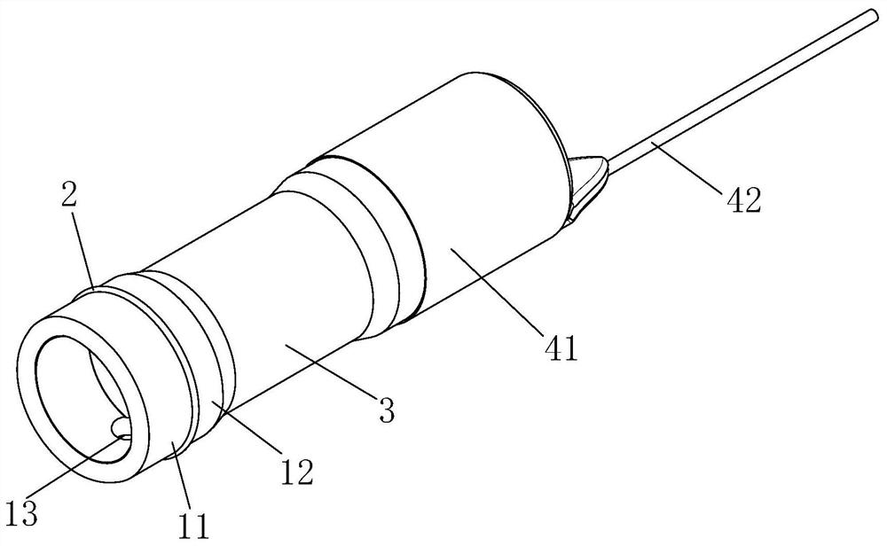
[0033] see Figure 1-4 , the present embodiment provides a hemostatic method used in esophageal varices surgery, the method uses a hemostatic device to stop bleeding from the injection hole of the vein in varicose vein surgery. The device is used together with a gastroscope in varicose vein surgery, and can press the bleeding point at the injection hole to avoid blood outflow. Wherein, the device includes a hemostatic sheath 1 , a limiting structure 2 , a connecting structure 3 and a varicose vein balloon 4 .

[0034] One hemostatic sheath is set on the probe front end of the gastroscope in the varicose vein operation. The hemostatic sheath 1 is a transparent structure made of polymeric polymer material, and the inner diameter of the hemostatic sheath 1 is smaller than the diameter of the probe to generate relative friction between the front end of the probe and the hemostatic sheath 1 . In this embodiment, the hemostatic sheath 1 includes a pressing section 11 and a sleeve ...

Embodiment 2

[0053] This embodiment provides a hemostatic method used in the operation of esophageal varices. The hemostatic device used in the hemostatic method is based on the embodiment 1 with an elastic hemostatic layer added. The elastic hemostatic layer is arranged on the outer wall of the hemostatic sheath 1, and it can be integrally formed with the hemostatic sheath 1, or it can be arranged separately. When the hemostatic sheath 1 performs hemostasis, the elastic hemostatic layer can just press against the bleeding point. Due to the elasticity of the elastic hemostatic layer, when the bleeding point is pressed, the elastic hemostatic layer will fully contact and adhere to the elastic tissue near the bleeding point to prevent blood from flowing out, thereby improving the sealing performance of hemostasis and improving the hemostatic effect. What needs to be explained here is that the elastic hemostatic layer can be made of transparent material, which is convenient for gastroscopy to...

Embodiment 3

[0055] This embodiment provides a hemostatic device used in the operation of esophageal varices. The hemostatic device includes a gastroscope and the hemostatic device used in Embodiment 1 or 2. Wherein, the hemostatic sheath 1 is set on the front end of the probe of the gastroscope, and the inner diameter of the hemostatic sheath 1 is smaller than the diameter of the probe so as to generate relative friction between the probe front end and the hemostatic sheath 1 . The limiting structure 2 is used to limit the end surface of the probe tip in the hemostatic sheath 1 so that the end surface of the probe tip and the end surface of the hemostatic sheath 1 are staggered. The connection structure 3 is set on the gastroscope. The 4 sets of varicose vein balloons are installed on the gastroscope. Among them, in the operation of varicose veins, after the foam sclerosant enters from the injection hole, the operator can observe and determine the corresponding bleeding point through the...


PUM


Abstract

The invention discloses an automatic hemostasis system used in the operation of esophageal varicose veins. The system includes a hemostatic device, an assembly mechanism, an insertion mechanism, an inflation mechanism, an injection mechanism and a control mechanism. The hemostatic device includes a hemostatic sheath, a limiting structure, a connecting structure and a varicose vein air bag. The insertion mechanism puts the gastroscope and the hemostatic device sleeved on the front end of the gastroscope into the esophagus. The inflation mechanism opens the varicose vein balloon, and inflates the varicose vein balloon to block the connection between the vein and the atrium to prevent the sclerosing agent in the varicose vein operation from flowing toward the atrium. The injection mechanism injects the foam hardening agent into the injection hole. The control mechanism controls the gastroscope to drive the movement of the hemostat. On the one hand, the present invention can prevent the outflowing blood from affecting the field of view of the gastroscope, facilitate the observation of the operation site, and ensure the success rate of the operation;

Description

technical field [0001] The invention relates to a blood vessel hemostatic device in the medical technology field, in particular to an automatic hemostasis system used in esophageal varicose vein surgery. Background technique [0002] Esophageal varices are now usually treated by injecting foam sclerosant. The foam sclerosant is punctured and injected through the operating hole of the gastroscope. After the injection of the foam sclerosant, the esophageal vein will leave an injection hole. The effect takes about five to ten minutes, but during this process, the injection hole will continue to bleed, which will affect the success rate of the operation on the one hand and make it difficult to observe the operation site. On the other hand, it will reduce the efficiency of the operation and is not conducive to foam sclerosing agents. hardening. Contents of the invention [0003] In order to solve the technical problem of injection hole bleeding in the existing varicose vein op...

Claims


Application Information

Patent Timeline
Patent Type & Authority Patents(China)
IPC IPC(8): A61B17/12A61B17/00
CPCA61B17/00234A61B17/12A61B17/12031A61B17/12109A61B17/12181A61B2017/00778A61B2017/00818A61B2017/12004A61B2017/1205
Inventor 孔德润庄小金彭杰杨思静宋绍方吴艾久
Owner 合肥中纳医学仪器有限公司
Why Eureka
Try Eureka