Method and device for automatic determination of the change of a hollow organ

A technology for automatically determining organs, applied in the direction of instruments, applications, instruments, etc. for radiological diagnosis, which can solve the problems of diagnosis, diagnosis failure, diagnosis delay, etc.

Pending Publication Date: 2021-03-12
SIEMENS HEALTHCARE GMBH
View PDF0 Cites 0 Cited by
  • Summary
  • Abstract
  • Description
  • Claims
  • Application Information

AI Technical Summary

Problems solved by technology

As a result, these changes cannot be diagnosed before complications arise, leading to delayed or failed diagnoses

Method used


Image

  • Method and device for automatic determination of the change of a hollow organ
  • Method and device for automatic determination of the change of a hollow organ
  • Method and device for automatic determination of the change of a hollow organ

Examples


Embodiment Construction

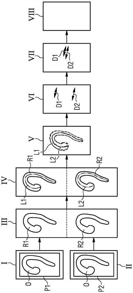
[0067] figure 1 Block diagram showing the process flow of a preferred method for automatically determining changes in a hollow organ O according to the present invention (see e.g. image 3 ).

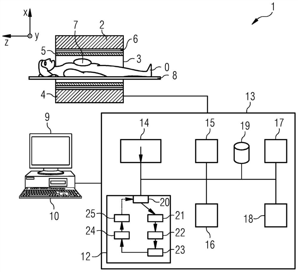
[0068] In step I, a first medical image P1 of an organ O is provided, already at a first point in time, for example with figure 2 The imaging device 1 shown records a first medical image P1.

[0069] In step II, a second medical image P2 of the organ O is provided, already at a (later) second point in time, for example also with e.g. figure 2 The imaging device 1 shown records a second medical image P2.

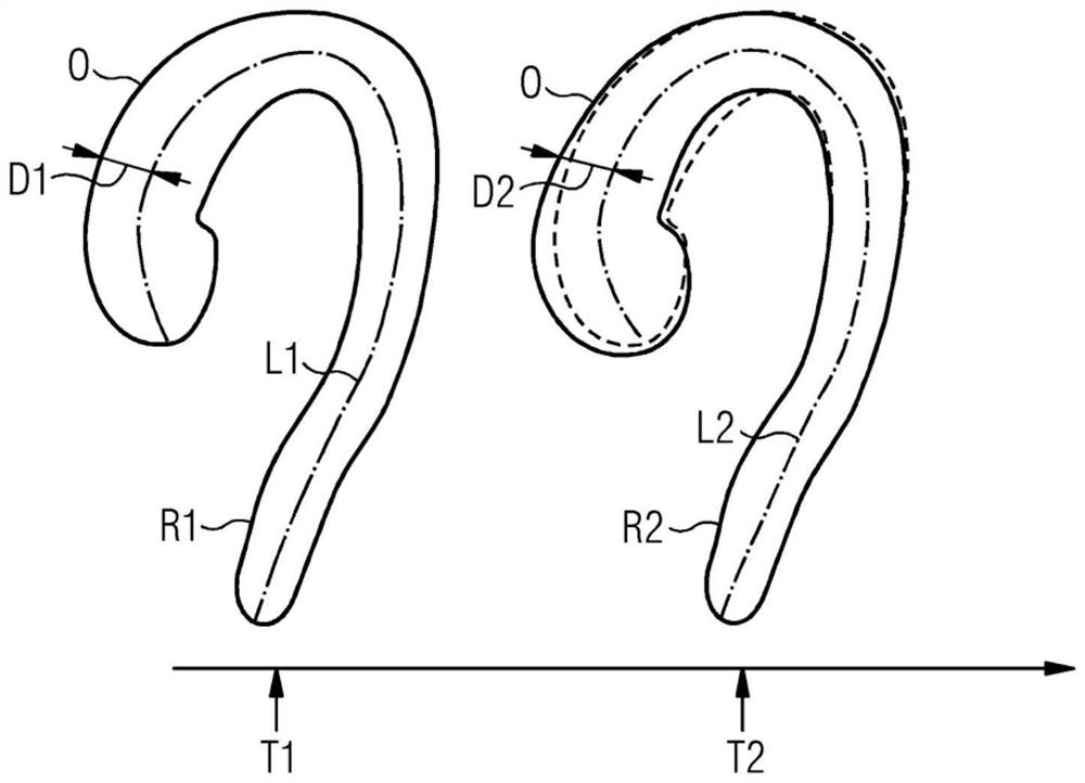
[0070] In step III, a first representation R1 of the organ O in the first image P1 and a second representation R2 of the organ O in the second image P2 are calculated. This can be performed in a single step or in two different (sub)steps.

[0071] In step IV, a first reference line L1 of the organ O is calculated based on the first representation R1 of the organ O, and a second ...


PUM


Abstract

The invention describes a method and a device for automatic determination of the change of a hollow organ. The method comprises the steps: providing a first medical image of the organ recorded at a first point of time, computing a first representation of the organ in the first image, computing a first reference-line of the organ based on the first representation of the organ, providing a second medical image of the organ recorded at a second point of time, computing a second representation of the organ in the second image, computing a second reference-line of the organ based on the second representation of the organ, registering of the first reference-line and the second reference-line to obtain a matching of the two representations of the organ and / or features derived from the representations, comparing the matched representations of the organ and / or the features derived from these representations. The invention further describes a related device, a related control device and a related medical imaging system.

Description

technical field [0001] The present invention describes methods and devices for automatically determining changes in hollow organs based on medical images. Background technique [0002] Dilation of vessels in humans is a serious risk to patients because these dilations - especially in the aorta - can progress towards an aneurysm which can burst or cause other adverse effects such as Heart attack, kidney damage, or stroke. Early detection of such dilatation is therefore crucial for many diseases, eg for the aorta, ie aortic dissection, aortic rupture and ruptured abdominal aortic aneurysm. Left untreated, these disorders often have serious and potentially fatal consequences. Therefore, it is important to monitor the progress of expansion of specific organs. [0003] Additionally, in the postoperative setting, for example, following endovascular aneurysm recovery, sac deflation is considered evidence of clinical success. The problem may also be prevalent in any hollow organ...

Claims


Application Information

Patent Timeline
Patent Type & Authority Applications(China)
IPC IPC(8): G06T7/00G06T7/33G06T7/10
CPCG06T7/0012G06T7/33G06T7/10G06T2207/10081G06T2207/10088G06T2207/10116G06T2207/30101G06T2207/20024G06T2207/30021G06T2207/10132G06T2207/30096G06T7/0016G06T2207/30061G06T2207/30172A61B6/504A61B6/032A61B6/486G06T7/11G06T7/337
Inventor 乔纳森·斯佩尔雷纳·克格尔
Owner SIEMENS HEALTHCARE GMBH
Why Eureka
Try Eureka