Intracranial blood vessel image registration method, electronic equipment and computer readable storage medium

An intracranial blood vessel and image registration technology, applied in the field of image processing, can solve the problems of uneven gray distribution, blurred medical images, various artifacts, etc., and achieve the effect of assisting the diagnosis of intracranial diseases

Inactive Publication Date: 2021-03-16
XIAN CREATION KEJI CO LTD
View PDF0 Cites 2 Cited by
  • Summary
  • Abstract
  • Description
  • Claims
  • Application Information

AI Technical Summary

Problems solved by technology

[0004] However, due to the complex tissue structure in the human body, the variety of organs, the adhesion between organs, or the movement of organs caused by the breathing movement of the human body, and the limitations of imaging equipment, medical images will appear blurred and the gray distribution is uneven. The edges of organs are not clear, and there are many artifacts. Secondly, the images of the same organ fr

Method used


Image

  • Intracranial blood vessel image registration method, electronic equipment and computer readable storage medium
  • Intracranial blood vessel image registration method, electronic equipment and computer readable storage medium
  • Intracranial blood vessel image registration method, electronic equipment and computer readable storage medium

Examples


Embodiment Construction

[0042] The present invention will be described in further detail below in conjunction with specific examples, but the embodiments of the present invention are not limited thereto.

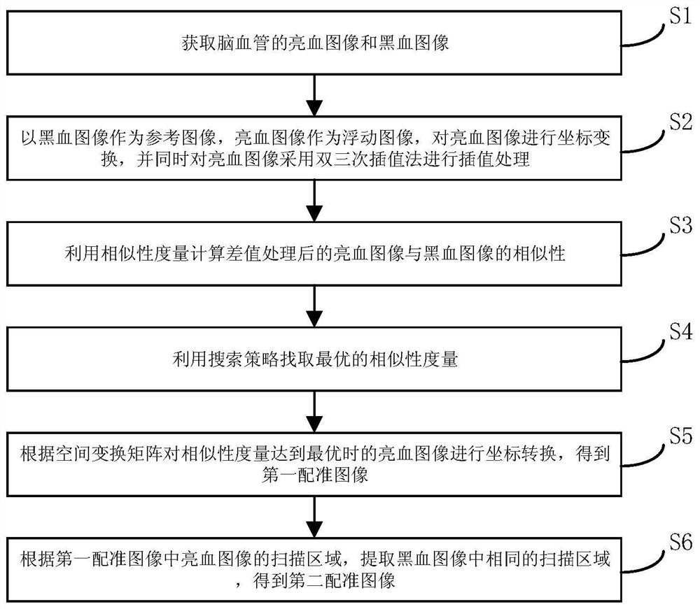
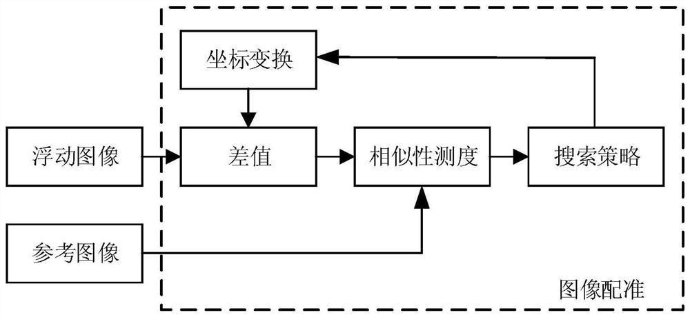
[0043] See figure 1 , figure 1 It is a flow chart of a method for intracranial blood vessel image registration provided by an embodiment of the present invention, as shown in figure 1 As shown, the intracranial blood vessel image registration method in the embodiment of the present invention includes:

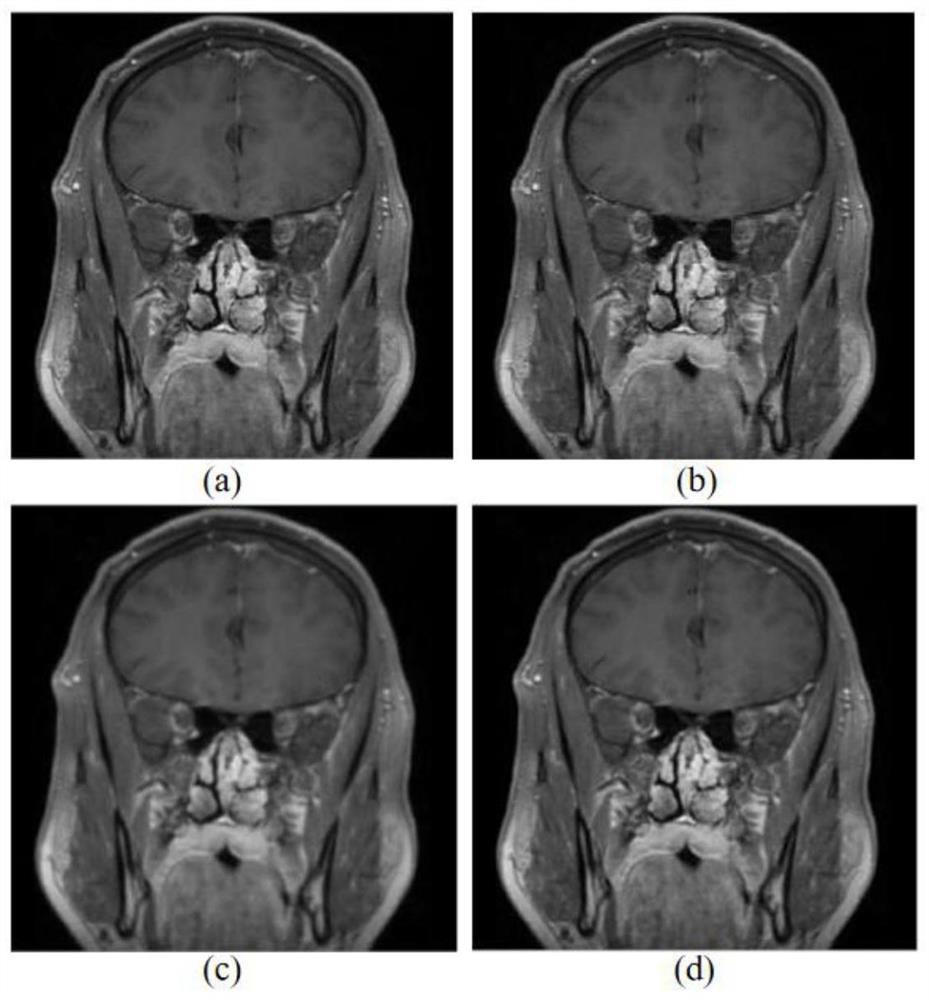
[0044] S1. Acquiring bright blood images and black blood images of intracranial blood vessels.

[0045] At present, methods based on lumen imaging are usually used to evaluate the degree of intracranial vascular lesions and vascular stenosis clinically, such as digital subtraction angiography (Digital Subtraction Angiography, DSA), CT angiography (Computed Tomography Angiography, CTA). ) and high-resolution magnetic resonance angiography (High-Resolution Magnetic Resonance Angiography, HRMRA). T...


PUM


Abstract

The invention discloses an intracranial blood vessel image registration method. The method comprises the following steps: acquiring a bright blood image and a black blood image of an intracranial blood vessel; taking the black blood image as a reference image and the bright blood image as a floating image, performing coordinate transformation on the bright blood image, and performing interpolationprocessing on the bright blood image by adopting a bicubic interpolation method; calculating the similarity between the bright blood image and the black blood image after interpolation processing byusing similarity measurement; finding out the optimal similarity measurement by utilizing a search strategy; according to the space transformation matrix, carrying out coordinate transformation on thebright blood image when similarity measurement is optimal, and obtaining a first registration image; and according to the scanning area of the bright blood image, extracting the same scanning area inthe black blood image to obtain a second registration image. According to the scheme, a doctor can be assisted in accurate intracranial disease diagnosis.

Description

technical field [0001] The technical field of image processing of the present invention specifically relates to a registration method for an intracranial blood vessel image, an electronic device, and a computer-readable storage medium. Background technique [0002] With the development of medical imaging technology, a variety of acquisition devices and imaging modes of medical images are emerging, which also brings the need for effective combination of medical images obtained from different medical devices or different acquisition modes of the same medical device. Promoted the development of medical image registration technology. [0003] Medical image registration technology can analyze the imaging results of the same patient from a single or multiple imaging modes. The registration images need to reflect different anatomical or functional information of the human body in the same coordinate system or on the same benchmark. There is no need for doctors to realize image spa...

Claims


Application Information

Patent Timeline
IPC IPC(8): G06T7/00G06T7/13G06T7/246G06T3/40G06K9/62G16H30/20
CPCG06T7/0012G06T7/13G06T7/248G06T3/4007G16H30/20G06T2207/30101G06F18/22
Inventor 石文
Owner XIAN CREATION KEJI CO LTD
Why Eureka
Try Eureka