
Intracranial blood vessel image flow void artifact elimination method and system

A technique of intracranial blood vessels and image flow, applied in the field of image processing, can solve the problems of complex realization, difficult blood vessel analysis, and inability to eliminate flow and void artifacts accurately and quickly, and achieve the effect of eliminating flow and void artifacts and good promotion.

Inactive Publication Date: 2021-03-16
XIAN CREATION KEJI CO LTD
View PDF0 Cites 0 Cited by
  • Summary
  • Abstract
  • Description
  • Claims
  • Application Information

AI Technical Summary

Problems solved by technology

Flow void artifacts will lead to poor description of blood vessels and surrounding tissues in black blood images, and will simulate wall thickening or plaque appearance of normal individuals, thereby affecting doctors' diagnosis and making blood vessel analysis extremely difficult
[0004] The current flow-space artifact elimination methods focus on improving medical image imaging technology, such as the adoption of new imaging principles, new pulse sequences, and research and development of new imaging modes, etc., but these methods are complex to implement and difficult to promote in clinical applications. Accurate and fast removal of flow void artifacts is not possible with devices without these pulse sequences or imaging modes

Method used


Image

  • Intracranial blood vessel image flow void artifact elimination method and system
  • Intracranial blood vessel image flow void artifact elimination method and system
  • Intracranial blood vessel image flow void artifact elimination method and system

Examples


Embodiment Construction

[0060] The present invention will be described in further detail below in conjunction with specific examples, but the embodiments of the present invention are not limited thereto.

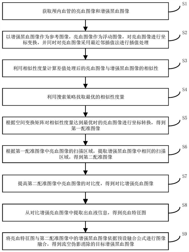
[0061] See figure 1 , figure 1 It is a flowchart of a method for eliminating flow space artifacts in intracranial blood vessel images provided by an embodiment of the present invention, as shown in figure 1 As shown, the method for eliminating flow space artifacts of intracranial blood vessel images in the embodiment of the present invention includes:

[0062] S1. Obtain bright blood images and enhanced black blood images of intracranial blood vessels.

[0063] At present, methods based on lumen imaging are usually used to evaluate the degree of intracranial vascular lesions and vascular stenosis clinically, such as digital subtraction angiography (Digital Subtraction Angiography, DSA), CT angiography (Computed Tomography Angiography, CTA). ) and high-resolution magnetic resonance angiography (H...


PUM


Abstract

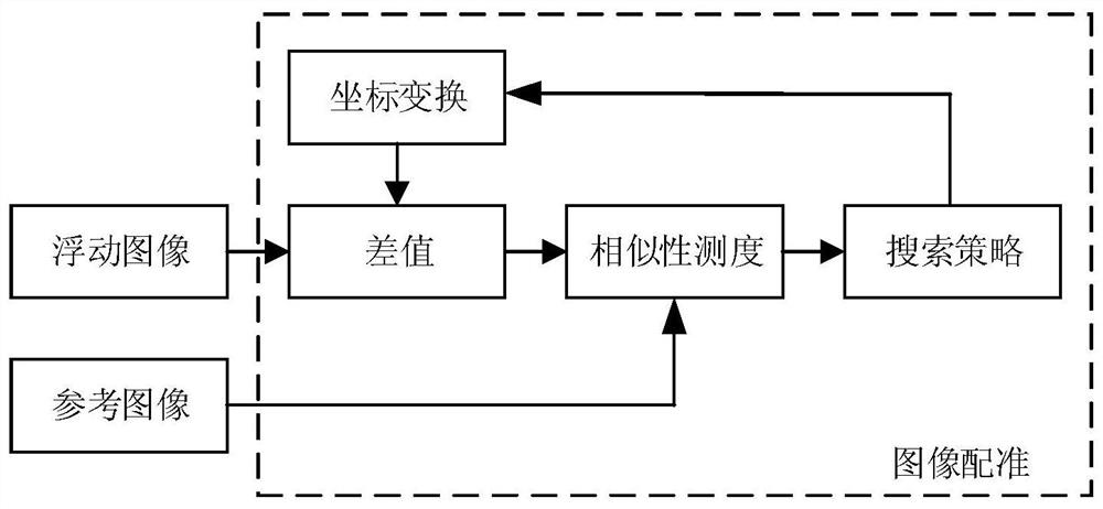
The invention discloses an intracranial blood vessel image flow void artifact elimination method. The method comprises the steps of acquiring a bright blood image and an enhanced black blood image ofan intracranial blood vessel; taking the enhanced black blood image as a reference image and the bright blood image as a floating image, performing coordinate transformation on the bright blood image,and performing interpolation processing on the bright blood image by adopting a nearest neighbor interpolation method; calculating the similarity between the bright blood image after interpolation processing and the enhanced black blood image by using similarity measurement; finding out the optimal similarity measurement by utilizing a search strategy; carrying out coordinate transformation on the bright blood image when the similarity measurement is optimal, and obtaining a first registration image; extracting the same scanning area in the enhanced black blood image according to the scanningarea of the bright blood image in the first registration image to obtain a second registration image; and carrying out flow void artifact elimination on the second registration image. According to the scheme of the invention, the flow void artifact can be quickly eliminated, more accurate and comprehensive blood vessel images can be displayed, and doctors can be assisted in accurate intracranialdisease diagnosis.

Description

technical field [0001] The technical field of image processing of the present invention, in particular, relates to a method and system for eliminating flow void artifacts in intracranial blood vessel images. Background technique [0002] According to the latest medical data, vascular diseases have seriously affected the life and health of contemporary people and become one of the diseases with a high mortality rate. Such as atherosclerosis, inflammatory vascular disease, vascular true neoplastic disease and so on. The common causes of vascular diseases are narrowing, blockage, rupture, and plaque in blood vessels. Clinically, high-resolution magnetic resonance angiography (High-resolution Magnetic Resonance Angiography, HRMRA) technology is generally used to detect the vessel wall and the degree of stenosis of the vessel, so as to provide doctors with the information required for diagnosis. [0003] Among them, the bright blood sequence in HRMRA can suppress the background...

Claims


Application Information

Patent Timeline
Patent Type & Authority Applications(China)
IPC IPC(8): G06T11/00G06T3/40G06T7/00G06T7/13G06T7/136G06T7/33G06K9/62G16H30/20
CPCG06T11/008G06T3/4007G06T7/0012G06T7/13G06T7/136G06T7/337G16H30/20G06T2207/30101G06T2211/424G06F18/22
Inventor 石文
Owner XIAN CREATION KEJI CO LTD
Learn More